5060
Magnetic Resonance Fingerprinting using phase cycling bSSFP (phc-MRF) in presence of B0 inhomogeneity.1Radiology Departiment, Case Western Reserve University, Cleveland, OH, United States
Synopsis
Magnetic Resonance Fingerprinting (MRF) is a newly developed approach for accurate and efficient multi parameter mapping. This work proposes a balanced steady state free precession (bSSFP) based MRF framework which introduces different phase cycling (phc-MRF) to compensate for banding artifact induced by B0 inhomogeneity. The phc-MRF was tested in both phantoms and in vivo. The preliminary results show the robustness of the phc-MRF to banding artifacts while maintaining the precision of parameter estimation and B0 sensitivity.
PURPOSE
Magnetic Resonance Fingerprinting1 is a newly developed approach for multiple tissue parameter mapping based on a single acquisition.This framework combines the transient state of the signal evolution with dictionary matching to achieve accurate and efficient multi-parameter maps. The original MRF design1 was based on a balanced steady state (bSSFP) acquisition which is sensitive to T1, T2 and off-resonance (B0) and provides high signal to noise ratio (SNR). The signal intensity is dependent on the off-resonance frequency, which allows B0 mapping but can also lead to banding artifacts in cases of significant B0 variations. A SSFP-MRF approach2 was proposed to eliminate banding artifacts, but it presents lower SNR and lack of B0 sensitivity.
The aim of this study is to exploit the flexibility of MRF to generate a bSSFP based approach which is unaffected by banding artifacts through cycling variation of the radiofrequency (RF) pulses phase (phc-MRF).
METHODS
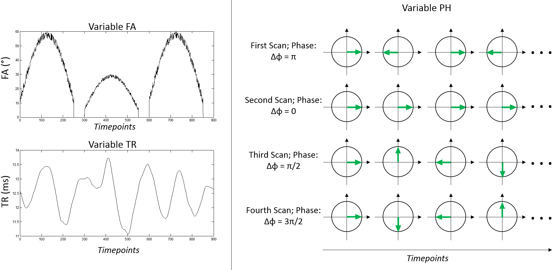
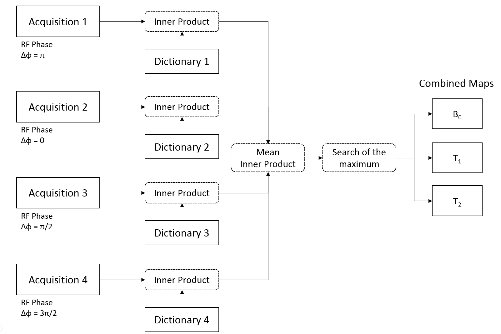
Based on prior work in phase-cycled bSSFP sequences3,4,5, our first proof-of-principle acquisition uses four bSSFP-MRF sequences collected one after each other. Each scan uses one arm of a variable density spiral6 trajectory for each TR to acquire an image (timepoint). For each of the 900 timepoints, the RF excitation angle (FA), the repetition time (TR), the RF pulse phase and the spiral arm orientation varies (Fig1). The four scans use a linear phase cycling with a phase increment (Δφ) for each timepoint of π, 0, π/2 and 3π/2 respectively (Fig1). Four different dictionaries are simulated for the four acquisitions. Each voxel is matched with the dictionaries through L2 normalized inner product (IP), then the IP values from all four matchings are averaged together to estimate T1, T2 and B0 maps (Fig2) selecting the atom providing the highest average IP.
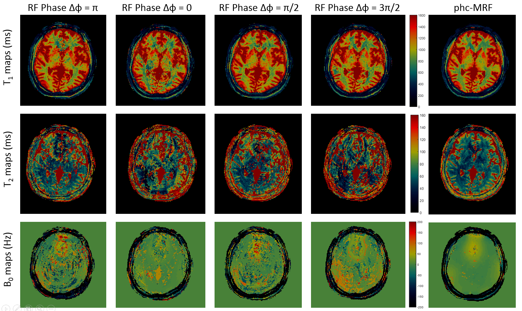
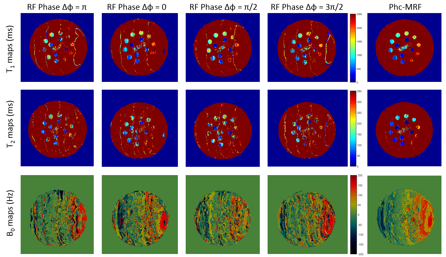
The phc-MRF has been tested on an healthy volunteer after informed consent in this IRB approved study and on an ISMRM/NIST MR system phantom7,8. The acquisition were performed on a 3T system (Skyra, Siemens, Germany) and data were acquired with a 16 channel array. Images of 256x256 base resolution and 300mm field of view were acquired. The volunteer acquisition was performed on a level of the brain close to the frontal sinus, where pronounced B0 inhomogeneity is present. The phantom acquisition was performed with a gradient applied along the x axis to generate a linearly varying frequency shift. The volunteer results were compared to the uncombined maps, while phantom results were compared to the values provided by NIST.
RESULTS
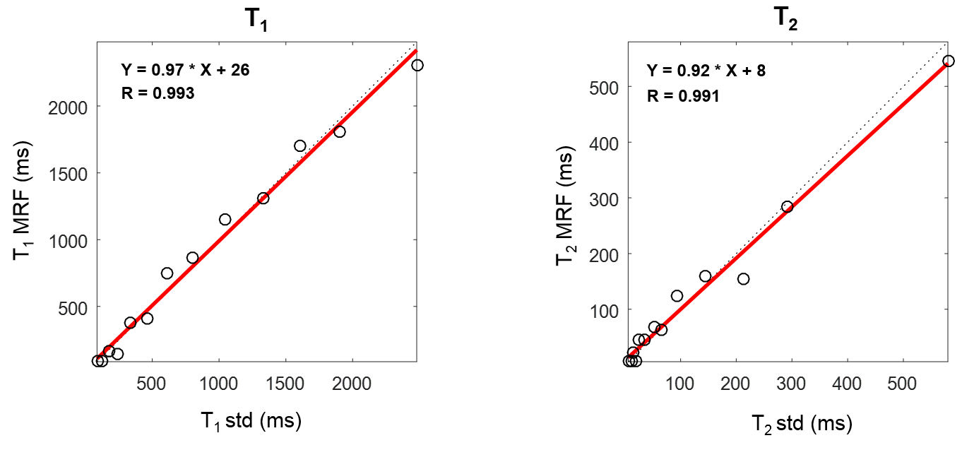
The T1, T2 and B0 maps obtained from the volunteer acquisition are shown in Figure 3. From the phc-MRF B0 maps (bottom right) one can notice the wide variation of off-resonance frequency due to the inhomogeneity induced by the tissue-air interface at the level of the sinus. Each of the single T1 maps presents artifacts in different region of the FOV, due to the different frequency response of each acquisition caused by the different RF pulses phase cycles. When combined in the reconstruction (rightmost column) nearly all banding artifacts are removed. The T2 maps of the individual acquisitions, instead, are of poor quality due to the artifacts and the limited number of timepoints used for reconstruction. When the four acquisition are combined, though, good quality T2 maps are obtained. The same maps obtained on the NIST phantom are shown in Figure 4. The inhomogeneity induced by the gradient caused extensive artifacts which were compensated by the phc-MRF which provided T1 and T2 values matching with the standard values within physiological range (Fig5).DISCUSSION
The results demonstrate that the MRF framework allows the combination of four different phase cycling to reduce the banding artifacts in a bSSFP-based acquisition. It has to be noted that each acquisition uses 900 timepoints, while the standard acquisition originally proposed1,2 acquires 3000 timepoints the total efficiency is minimally affected.
As future works, further characterization of the approach will be performed, the four acquisitions will be merged in a single one and the phc-MRF performances in other regions of the body will be investigated. We believe that the method will be useful in areas where the wide B0 inhomogeneity along the extended field of view makes the collection of bSSFP based images challenging due to the persistent presence of banding artifacts.
CONCLUSION
In conclusion this work presents a proof of principle implementation of phc-MRF, which is capable of compensating for banding artifacts. This a first fundamental step towards an MRF acquisition suitable for all body applications where a bSSFP based approach would provide high SNR and ability to map tissue parameters and static field inhomogeneity.Acknowledgements
The authors would like to acknowledge funding from Siemens Healthcare and NIH grants NIH 1R01EB016728 and NIH 5R01EB017219.References
[1] Dan Ma et al,”Magnetic Resonance Fingerprinting” Nature 2013; 495(7440):187
[2] Yun Jiang et al,”MR fingerprinting using fast imaging with steady state precession (FISP) with spiral readout” MRM 2015; 74(6):1621
[3] Neal Bangerter et al, “Analysis of multiple-acquisition SSFP”, MRM 2004; 51(5):1038
[4] Marcus Björk et al, “Parameter Estimation Approach to Banding Artifact Reduction in Balanced Steady-State Free Precession”,MRM 2014; 72(3):880
[5] Thomas Benkert et al, “Dynamically Phase-Cycled Radial Balanced SSFP Imaging for Efficient Banding Removal”, MRM 2015; 73(1):182
[6] Jin H. Lee et al,”Fast 3D imaging using variable-density spiral trajectories with applications to limb perfusion”, MRM 2003; 50(6):1276
[7] Russek SE, et al. “Characterization of NIST/ISMRM MRI System Phantom”. Proceedings of the 20th meeting of the ISMRM, Melbourne, Australia, 2012
[8] Kathryn E Keenan et al,”Multi-site, multi-vendor comparison of T1 measurement using ISMRM/NIST system phantom”, Proceedings of the 24th meeting of the ISMRM, Singapore, 2016. Abstract #3290
Figures