5055
A Method of Fat Saturation with Better Transmit Field Inhomogeneity Immunity1MR Research China, GE Healthcare, Beijing, People's Republic of China, 2MR Engineering, GE Healthcare, Beijing, People's Republic of China
Synopsis
Discrimination between signals from fat and water is of great importance in clinical practice. Chemical saturation, the most widely used fat suppression method, has degraded performance in an in-homogeneity transmit field, and may jeopardize the diagnosis effectiveness. In this abstract, we proposed an improved fat saturation method employing multiple chemical saturation segments. Phantom and volunteer evaluation results show that new method has much less residual lipid signal and can achieve more uniform lipid saturation over big field of view under inhomogeneity condition. Which support the conclusion that proposed method has superior transmit field inhomogeneity immunity.
Introduction
Lipid presented as marked hyperintense both on T1 and T2-weighted images in MR scan. Clinically, it is often desirable to suppress the lipid signal. Especially for those lipid-rich anatomies’ examinations. For example, in breast scan, good fat saturation is important to delimit the lesions. Chemical shift selective (CHESS) radiofrequency pulse is commonly used to suppress lipid signal. However, the performance of CHESS pulse is highly depended on the uniformity of both excitation RF field (B1) and static magnetic field (B0). Especially when there is B1-inhomogeneity exist, the CHESS’s performance is quite limited in effectiveness without parallel RF transmitting capability. Adiabatic chemical saturation pulse [1] has very promising performance under B1-inhomo condition, but its long pulse duration time as well as high Specific Absorption Rate (SAR) are the main concerns. Dixon fat/water imaging based on 2 or 3 echoes is another approach [2]. However, it takes significantly longer scan time to acquire additional echoes.
In this study, we evaluated the feasibility of achieving fat saturation with an improved B1-inhomogeneity immunity by combining multiple CHESS chemical saturation (CS) pulse segments.
Method
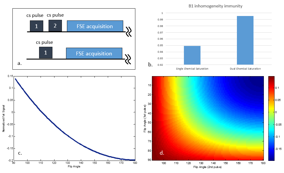
Unlike traditional fat saturation, which uses single CS pulse, what we proposed, termed as Dual Chemical Saturation (DCS), employs 2 successive CS pulses. Figure 1-a shows the pulse sequence diagram of traditional CHESS and DCS. For traditional CHESS approach, given a set of acquisition parameters, the flip angle for CHESS pulse to null lipid signal can be derived by solving Bloch equation. As the plot depicted in Figure 1-c, the normalized fat signal is expected to be nulled with a flip angle of 110 degree. For DCS, combining different flip angles for 1st and 2nd chemical saturation pulses, Bloch calculation will generate one “image” with normalized fat signal indexed by flip angle values of the 2 CS pulses. Figure 1-d shows the normalized fat signal “image” and demonstrated the key difference of DCS compared with CHESS: DCS has multiple fat signal nulling points with different B1-inhomogeneity immunity.
The B1-inhomogeneity immunity (BI) is evaluated by:
BI = [(1 – |NF-0.5dB|)+(1 – |NF|)+(1 – |NF+0.5dB|)]/3
Where NF is the normalized fat signal and the subscript denotes the B1 field in-homogeneity in dB. Final flip angles for DCS is determined by choosing the nulling point with maximum BI. Figure 1-b compared calculated BI of CHESS and DCS with final flip angles. DCS shows superior performance in fat saturation under B1-inhomogeneity condition.
Phantom and Volunteer Evaluation
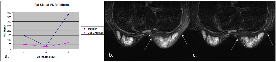
Phantom evaluation was done with a GE 3.0T MR Scanner (Discovery 750; GE Medical system, Milwaukee, WI) and a vegetable oil phantom. T2 weighted fast spin echo (FSE) with fat saturation of CHESS and DCS techniques were scanned under normal and B1-inhomogeneity conditions. We mimicked B1-inhomogeneity by manually adjusting transmit amplifier’s gain to achieve ±1dB attenuation of the B1 field. Fat signal was measured with ROI on the phantom and summarized in Figure 2-a. DCS achieved 67% and 84% lower fat signal under -1dB and +1dB condition respectively. In normal condition, the 2 techniques have similar performance.
After the informed consent was obtained, a female healthy volunteer was recruited for in-vivo evaluation. CHESS and DCS performance was compared in a bilateral breast T2 FSE scan. The scan parameters are: axial plane, 36cm FOV, 5.0mm slice thickness, TR 5000ms, TE 85ms, echo train length 16, #slice 19, number of average 2. One of the slices is shown in Figure 2.b~c. Residual lipid signal was found in the right breast (arrow) from CHESS image, while fat signal in DCS image is obviously lower and the left/right breast appears much more even compared with CHESS.
Discussion
Adding an additional CS pulse segment to scan will double the time needed for lipid saturation. The final impact on scan time depends on the duty cycle of fat saturation segment on the whole sequence. For the most commonly used FSE, DCS will add 2 – 4 seconds to the total scan time. Beside of bilateral breast, DCS is expected to show improvement on other large FOV scan. For example, abdomen, pelvis and spine. The implementation in this abstract uses 2 chemical saturation pulses. However, this technique has the potential extension to more CS pulses, to achieve even better B1 inhomogeneity immunity. And the cost, of course, would be even longer scan time.Conclusion
By combining 2 chemical saturation pulses, DCS achieved better B1-inhomogeneity immunity compared with traditional CHESS pulse. Less residual lipid signal and more uniform fat saturation will benefit radiologist as well as patients from a less likely compromised diagnosis.Acknowledgements
No acknowledgement found.References
1. Daniel Rosenfeld, Design of adiabatic pulses for fat-suppression using analytic solutions of the bloch equation, MRM Volume 37 Issue 5, Page 793-801.
2. Reeder SB, McKenzie CA, Pineda AR, Yu H, Shimakawa A,Brau AC, et al. Water-fat separation with IDEAL gradient echo imaging. J Magn Reson Imaging.2007;25(3):644-652.
Figures