5054
Improved image-based navigators (iNAVs) for free-breathing cine DENSE using principle component analysis to separate the stimulated echo and T1 relaxation signals1Biomedical Engineering, University of Virginia, Charlottesville, VA, United States, 2MR R&D Collaborations, Siemens Healthcare, Atlanta, GA, United States, 3Department of Radiology and Imaging Sciences, Emory University, Atlanta, GA, 4Electrical and Computer Engineering, University of Virginia, Charlottesville, VA, United States, 5Radiology, University of Virginia, 6Medicine, University of Virginia
Synopsis
Cine displacement encoding with stimulated echoes (DENSE) is an accurate strain imaging technique that generally requires breath-holding. We have recently developed a free-breathing method with image-based navigators (iNAVs) that makes use of the localized signal generation property of stimulated echoes (STE) to facilitate the estimation of heart motion due to respiration. However the non-localized echo due to T1 relaxation presents challenges. We propose to use principle component analysis (PCA) to separate the STE and T1-relaxation echo signals and improve the accuracy of motion estimation with iNAVs for free-breathing cine DENSE.
Background
Myocardial strain imaging is increasingly used for the assessment of cardiac function. Cine displacement encoding with stimulated echoes (DENSE)1 is an accurate and reproducible strain MRI method, but generally requires breath-holding, which is difficult for many heart disease patients. We have recently developed a free-breathing spiral cine DENSE method with image-based navigators (iNAVs) that makes use of the localized signal generation property of stimulated echoes (STE) to facilitate the estimation of heart motion due to respiration,2 however a non-localized and interfering echo due to T1 relaxation (predominantly from fat) presents challenges. While we previously used a k-space filter (as in HARP MRI)3 to suppress the T1 relaxation echo, we found that this method has limitations and that the T1 relaxation echo can still interfere with the heart-localized iNAVs and reduce the accuracy of motion estimation. Accordingly, we propose to use principle component analysis (PCA) to separate the STE and T1 relaxation echo and improve the accuracy of iNAV motion estimation for free-breathing cine DENSE.Methods
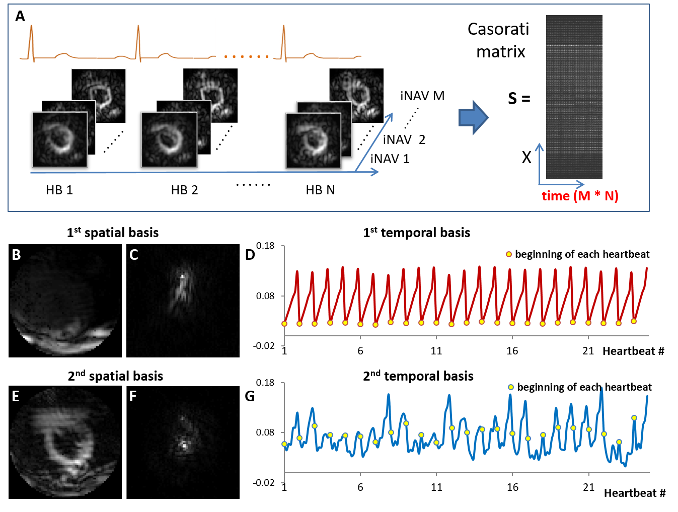
A cine DENSE sequence was modified to use variable density spirals, golden angle rotations, and localized signal generation of the STE (using 3 orthogonal slice-selective RF pulses). Volunteers (n=6) were scanned using a 3T system (Trio, Siemens). Mid-ventricular short-axis slices were acquired during free-breathing using: field-of-view of 160-200 mm, 6 interleaves per image, 2 interleaves per heartbeat, temporal resolution of 30 ms, matrix size of 70x70, 2-point phase-cycling, and 4 averages, with a total scan time of 72 heartbeats. We investigated the use of PCA in order to separate the STE and T1 relaxation signal, and subtract the T1 relaxation signal. Using the same raw data, we performed iNAV processing and estimation of respiratory motion using three different methods: (a) manual tracing of the heart using iNAVs computed with k-space filtering of the T1 relaxation echo as a reference standard, (b) 2D cross-correlation applied to iNAVs computed with k-space filtering of the T1 relaxation echo, and (c) 2D cross-correlation applied to iNAVs with PCA filtering.
The PCA-based filtering method is shown in Figure 1. As shown, initial iNAV images are reconstructed by combining 8 consecutively acquired spiral interleaves, resulting in multiple iNAVs per heartbeat for all heartbeats of the scan. All initial iNAVs including both phase-cycling dimensions are reorganized into a Casorati matrix (spatial-temporal) (Figure 1 (A)). PCA is performed to decompose the Casorati matrix into spatial and temporal bases. The first spatial and temporal bases are found to correspond predominantly to the T1 relaxation echo (Figure 1 (B, C)), and the second to the STE (Figure 1 (E, F)). Thus, the first spatial and temporal basis can be removed to reduce T1 relaxation signals (PCA-based filtering). Motion estimation using iNAVs before and after PCA filtering was compared to manual tracing by calculating root-mean-square error (RMSE) of the translations. The Wilcoxon signed rank test was used for statistical analysis.Results
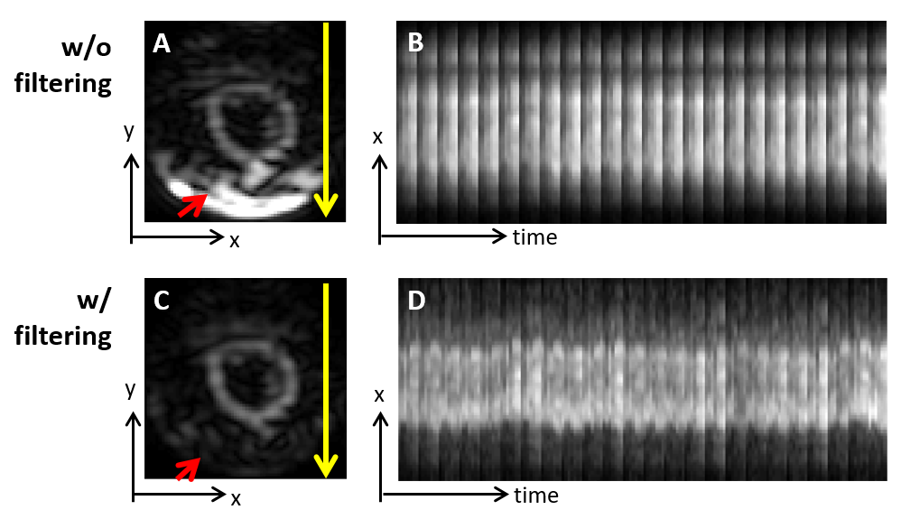
Example iNAVs without and with PCA-based filtering are shown in Figure 2 (A, C). PCA-based filtering removed the strong signal outside the heart due to T1 relaxation (red arrow). The temporal image series are collapsed into 1D projections in the x-direction and are shown in Figure 2 (B, D). With PCA-based filtering, respiratory motion is better visualized compared to without PCA-based filtering.
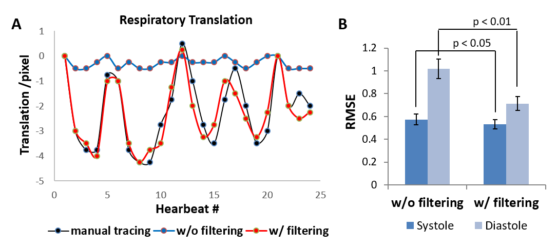
An example of respiratory-induced translations estimated from a set of diastolic iNAVs is shown in Figure 3 (A). Respiratory motion estimated from iNAVs with PCA-based filtering closely matches manual tracking while the motion estimated from iNAVs without PCA-based filtering is not accurate. Figure 3B summarizes the RMSE of translation estimation for all subjects and shows that for both systolic and diastolic iNAVs, the RMSE with PCA-based filtering is significantly lower than that without filtering (systole, 0.56 +/- 0.06 vs. 0.63 +/- 0.07, p < 0.05; diastole, 0.71 +/- 0.09 vs. 1.02 +/- 0.12, p < 0.01). The improvement in motion estimation in diastole is greater than in systole.
Conclusion
The application of PCA to cine DENSE iNAVS successfully separates the STE and T1 relaxation signals, and PCA-based filtering significantly improves the quality of localized STE iNAVs and the accuracy of respiratory motion tracking. Future work will translate the improved iNAV methods to improved free-breathing cine DENSE myocardial strain imaging.Acknowledgements
This work was supported by NIH R01EB001763 and an award from the American Heart Association and The Children’s Heart Foundation, AHA16PRE30680001.References
1. Kim, Daniel, et al. "Myocardial Tissue Tracking with Two-dimensional Cine Displacement-encoded MR Imaging: Development and Initial Evaluation." Radiology 230.3 (2004): 862-871.
2. Cai, Xiaoying, et al. “Free-breathing 2D Cine DENSE with localized excitation, self-navigation and motion correction”. Journal of Cardiaovascular Magnetic Resonance 2016, 18(Suppl 1):P319
3. Osman, Nael F., et al. "Cardiac motion tracking using CINE harmonic phase (HARP) magnetic resonance imaging." Magnetic resonance in medicine: official journal of the Society of Magnetic Resonance in Medicine/Society of Magnetic Resonance in Medicine 42.6 (1999): 1048.
Figures