5052
Magnetic Resonance Imaging under Highly Inhomogeneous B0 Fields using Missing-Pulse Steady-State Free Precession (MP-SSFP)1Center for Magnetic Resonance Research,Department of Radiology, University of Minnesota, Minneapolis, MN, United States
Synopsis
In vivo human brain imaging under highly inhomogeneous B0 field of 250 kHz off-resonance variation over 20 cm is demonstrated. The B0 field inhomogeneity was generated by mounting a head gradient coil at 36 cm off the isocenter of a 90-cm 4T magnet. Brain imaging was performed with 3D missing-pulse steady-state free precession using the inhomogeneous field gradient for spatial (readout) encoding. Frequency-modulated pulses were employed to excite the widely distributed spin frequencies in the inhomogeneous field with easily achievable RF peak power (~1 kW). By providing combined T1, T2, and high diffusion weighting, images with clear delineation of brain anatomy were produced.
Purpose
Currently used standard MRI systems require highly homogeneous magnetic fields, in which the Larmor frequency variation is on the order of only one or two hundred hertz. Here we introduce an MRI method using missing-pulse steady-state free precession (MP-SSFP)1 that is capable of producing good quality images under a permanent B0 field inhomogeneity of roughly 250 kHz over 20 cm.Methods
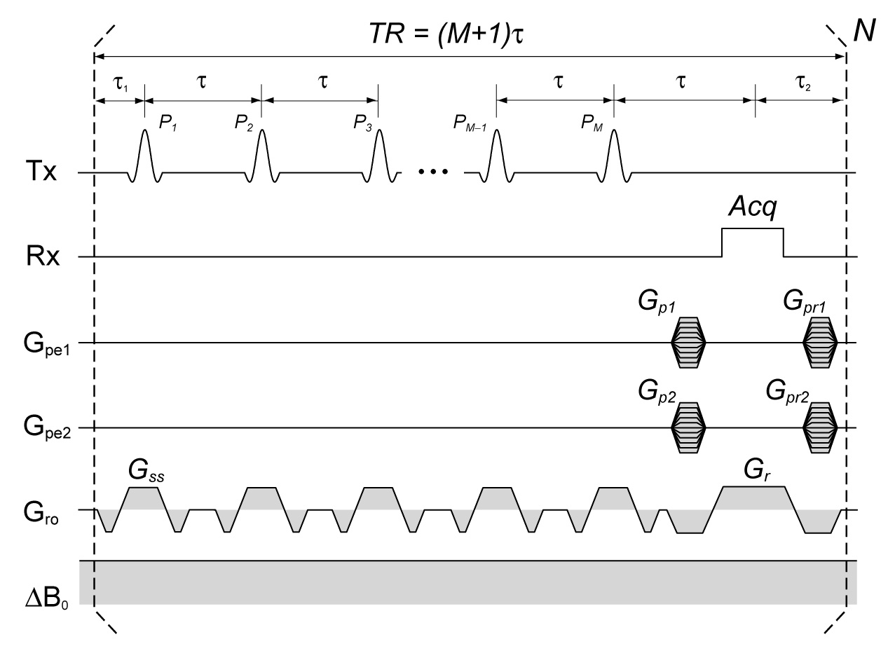
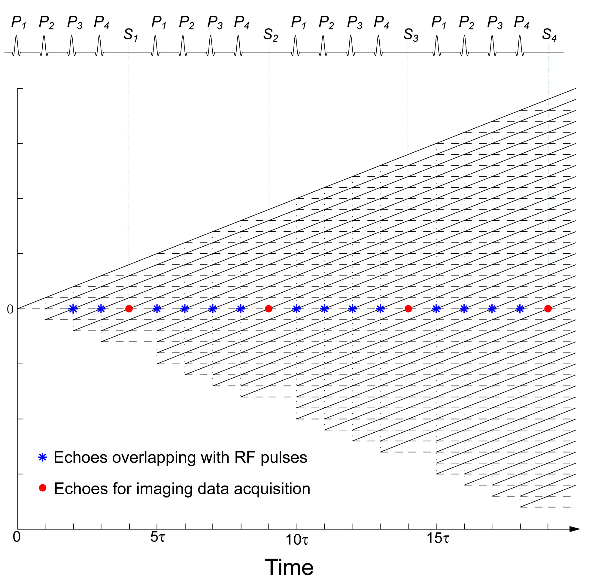
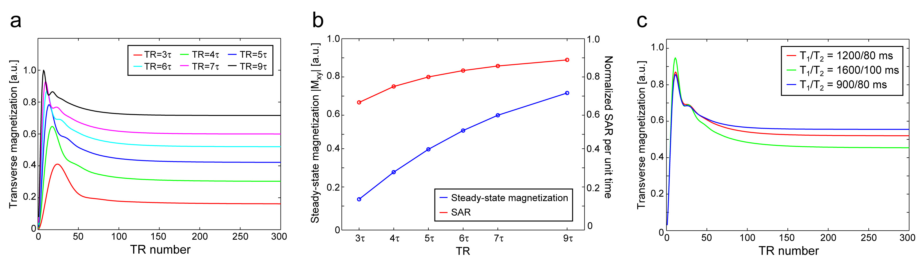
A permanently inhomogeneous static field was introduced by mounting a head gradient coil at 36 cm away from the magnet center of a 4T human MRI system (Agilent Technologies, Inc.), which resulted in approximately 250 kHz off-resonance variation over 20 cm. To preserve spin signals under the highly inhomogeneous field, a 3-dimensional MP-SSFP sequence was employed (Fig.1), where echoes consisting of overlapping spin and stimulated echoes refocus at the missing pulse timings regardless of the field inhomogeneity (Fig.2). The permanent B0 field gradients mostly along the z direction were used as spatial encoding (readout) gradients. For RF excitation, a frequency-modulated (FM) pulse that is a stretched hyperbolic secant pulse (HS2)2 was employed to cover the wide frequency range generated by the inhomogeneous field (>100 kHz). The dependence of the steady-state magnetization on the missing pulse interval was investigated using simulations, and the transverse magnetization reaching the steady-state was calculated from a phase diagram3 using the following parameters: pulse interval (τ)=3 ms, flip angle (FA)=4°, and T1/T2=1200/80 ms. To study the dependence of the steady-state magnetization on T1 and T2 relaxation, simulations were performed using one missing pulse in every 6 pulses (TR=6τ) for three T1 and T2 combinations (T1/T2=1200/80, 1600/100 and 900/80 ms).
Under the inhomogeneous B0 field, in vivo human brain imaging was performed with the following sequence parameters: τ=2.6 ms, pulse missing is every 6 pulses (TR=15.6 ms), HS2 pulse excitation with FA=4°, BW=140 kHz and Tp=700 μs (corresponding to a time-bandwidth product (TBP)=100), readout bandwidth = 180 kHz, cylindrical k-space sampling, and scan time = 5.5 min. Pulsed slab-selective and readout gradients, Gss and Gr, were applied to mitigate a portion of the B0 inhomogeneity during excitation and acquisition. SAR was within the FDA guideline. A quadrature TEM coil was used for transmit and receive. Since the field gradient associated with the inhomogeneous B0 field was not linear, a reconstructed image showed image distortion along the z direction. The image distortion was corrected based on a measured B0 field map. Image reconstruction was performed with a reconstruction routine written with C++ and image distortion correction was done with Insight Toolkit (ITK)4.
Results
The simulation shows a rapidly increasing amplitude of the refocused transverse magnetization at the beginning of the MP-SSFP sequence, that reaches a maximum and then slowly decreases to a steady-state level (Fig.3a). In addition, the steady-state transverse magnetization increases as the missing pulse interval becomes longer (i.e. longer TR). SAR per unit time also increases gradually as the missing pulse interval increases, but the steady-state magnetization increases at a faster rate (Fig.3b). One drawback of using longer missing pulse interval is an increase of total scan time due to longer TR. The steady-state magnetization attains different steady-state magnetization levels, depending on T1 and T2 values (Fig.3c), which generate contrasts in images.
A brain image reconstructed with inverse Fourier transformation showed significant image distortion resulting from the nonlinear inhomogeneous B0 field (Fig.4a,b). Distortion correction using a measured B0 field map nicely eliminated the image distortion (Fig.4c-f).
Discussion
The proposed 3D MP-SSFP technique uses the permanent inhomogeneous B0 field gradient as a frequency encoding (readout) gradient. The nonlinear inhomogeneous gradient field generates not only image distortion, but also spatially varying diffusion weighting in reconstructed images. There was a gradual signal drop toward the top of the brain because of the steeper B0 field gradient (Fig.4a-c). In this study, relatively short τ (2.6 ms) was used to avoid excessively strong diffusion weighting in those regions.
Signal excitation with broad bandwidth (>100 kHz) is another challenging aspect of MRI under highly inhomogeneous fields, which makes conventional spin-echo sequences using 180 degree refocusing pulses impractical. The achievable excitation bandwidth is a severe limitation for conventional amplitude modulated (AM) pulses. Specifically, the required pulse width of common AM pulses (e.g., sinc and Gaussian) is <50 μs for 100 kHz bandwidth, which may require extremely high RF peak power even for low flip angles. FM pulses used in this study can significantly alleviate the peak power demand by using high TBP pulses.
Conclusions
3D MP-SSFP can achieve in vivo human brain imaging under an inhomogeneous B0 field of 250 kHz off-resonance over 20 cm.Acknowledgements
This study was supported by NIH grants R24MH105998, P41EB015894, S10RR023730 and S10RR027290.References
1. Patz, S., S.T. Wong, and M.S. Roos, Missing pulse steady-state free precession. Magn Reson Med, 1989. 10(2): p. 194-209.
2. Garwood, M. and L. DelaBarre, The return of the frequency sweep: designing adiabatic pulses for contemporary NMR. J Magn Reson, 2001. 153(2): p. 155-77.
3. Scheffler, K., A pictorial description of steady-states in rapid magnetic resonance imaging. Concepts in Magnetic Resonance, 1999. 11(5): p. 291-304.
4. Yoo, T.S., et al., Engineering and algorithm design for an image processing Api: a technical report on ITK--the Insight Toolkit. Stud Health Technol Inform, 2002. 85: p. 586-92.
Figures