5041
Omitting the control condition using self-control super-selective Arterial Spin Labeling to reduce total scan time for flow territory mapping1Clinic for Radiology and Neuroradiology, University Hospital Schleswig-Holstein, Kiel, Germany, 2Tomographic Imaging Department, Philips Research, Hamburg, Germany
Synopsis
Selective Arterial Spin Labeling (ASL) is established to perform non-contrast enhanced flow territory mapping. In super-selective pCASL only one artery of interest is labeled while the efficiency in contralateral arteries is near zero. To obtain a holistic picture of all brain perfusion territories, the label and control experiments have to be repeated for each artery, prolonging scan time. In this study, it is hypothesized that due to the (almost) negligible signal contribution of non-tagged arteries, selective perfusion images can be calculated from a single scan that is performed without the acquisition of control images.
Introduction
Arterial Spin Labeling (ASL) is an established technique to perform non-contrast enhanced MR perfusion imaging. Modifications of the labeling module allow for the selective visualization of individual brain feeding arteries, e.g. the internal carotid arteries (ICAs) and vertebral arteries (VAs). Generally, ASL approaches rely on two acquisitions, namely a “label” (inversion) and a “control” (without inversion) condition, which are subsequently subtracted to remove the static tissue signal, yielding perfusion weighted images. In super-selective pCASL only a single artery of interest is labeled while the labeling efficiency in contralateral arteries is almost zero [1]. To obtain a holistic picture of brain perfusion using super-selective tagging, the label and control experiment have to be repeated for each selectively labeled artery, prolonging total scan time. In the present study, it is hypothesized that due to the (almost negligible) signal contribution of non-tagged arteries, selective perfusion images can be calculated without an additionally acquired control image by using the tag images only.Materials and Methods
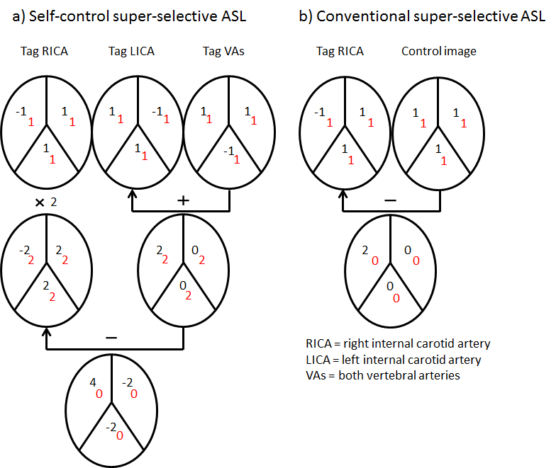
Six healthy volunteers underwent MR scanning under the general protocol for sequence development, approved by the local ethical committee. Imaging was performed on a Philips 3T Achieva (Philips, Best, The Netherlands) scanner using a standard 32 channel SENSE Head coil. ASL parameters were: 1800ms Label and 2000ms post labeling delay with background suppression pulses at 1850ms and 3250ms as recommended in [2]. Image acquisition consisted of a multislice 2D EPI readout with 2.3*2.3*4mm voxel size covering 16 slices with a gap of 1.6mm. Planning of the individual labeling foci for all selected arteries was performed on a low-resolution time-of-flight scan positioned across the neck. During scanning, the individual labeling focus positions for each vessel were cycled periodically so that all selective data acquisitions were performed within a single measurement. For each of the major arteries (right and left ICA and VAs), 20 images were acquired, resulting in a total acquisition time of 4:13min. As a comparison, conventional super-selective ASL with a separate label and control image was performed with the same parameters and took 8:26 min in total. Non-selective ASL imaging took 3:06 min. In both of the latter methods, perfusion weighted images were obtained by subtraction of label and control images. In the presented approach, the flow territories were calculated by combining the images of the contralateral arteries and subtracting the image of the tagged artery twice (Figure 1a). The resulting negative signal of the contralateral arteries is removed via logical indexing. In order to obtain quantitative information from the perfusion images, the algorithm presented in [2] was used, after the images were divided by the square root of 2 to correct for the change in signal-to-noise ratio resulting from the combination of multiple images [3, 4]. All calculations were performed using Matlab R2013a (The Mathworks, Natick, MA).Results and Discussion
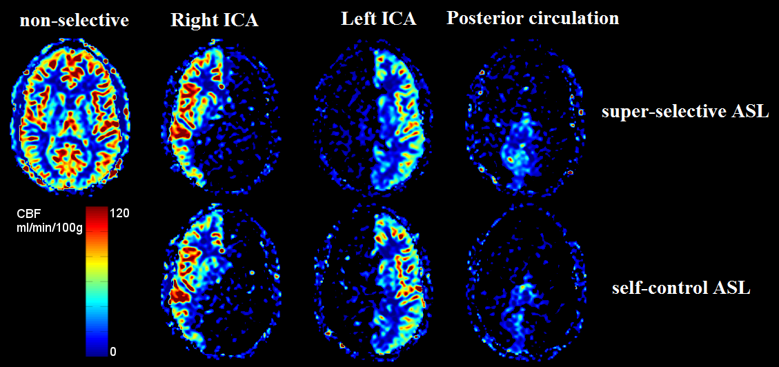
Images of all individual perfusion territories could be obtained. The resulting quantitative images are in concordance with conventional super-selective as well as with non-selective ASL (Figure 2). To acquire selective perfusion territories using super-selective ASL, at least two image acquisitions are needed. Generally, this is achieved by acquiring a label image with a matching control image. However, the need for acquiring two corresponding images increases scan time and makes the acquisition prone to patient motion. The presented method demonstrates that the information of the contralateral (non-tagged) arteries can be used for calculating selective perfusion territories instead of using an additionally acquired control image when three individual arteries are labeled. In cerebrovascular diseases, however, the flow territories of additional intracranial arteries need to be visualized selectively. It is possible to use super-selective ASL to label such arteries, which will require increasing the number of tagging acquisitions as well as adapting the decoding scheme. Such approach is subject to further investigations. As this calculation assumes perfect inversion within a single artery and zero inversion outside, the actual effects of the varying labeling efficiency in contralateral arteries have to be further investigated as well.Conclusion
In this study, a method to post-process super-selective ASL images using only the label data is presented which makes it possible to generate perfusion weighted images of individual flow territories without the necessity to acquire additional control images. Using this approach, flow territory mapping can be performed in shorter scan times than using conventional super-selective ASL.Acknowledgements
No acknowledgement found.References
[1] Helle M et. al. Magn Reson Med 2010;64:777-86
[2] Alsop D et. al. Magn Reson Med 2014;73:102-16
[3] Günther M. Magn Reson Med 2006;56:671-5
[4] Gudbjartsson H and Patz S. Magn Reson Med 1995;34:910-4
Figures