5038
DTI measurements with exceptional resilience to field heterogeneities in challenging brain regions1chemical Physics, Weizmann Institute of Science, Rehovot, Israel
Synopsis
This study presents a new diffusion tool based on a novel single-shot 2D MRI method called xSPEN. xSPEN is characterized with unusual resilience to field heterogeneities, but extending it to DTI requires to overcome the strong intrinsic diffusion weighting of this technique. To achieve this we formulated xSPEN’s diffusion weighting using a novel, spatially localized b- matrix analysis, and devised a novel diffusion-weighting scheme that overcomes xSPEN’s original limitations. These methods were numerically validated and applied to new DTI xSPEN sequences with which we mapped diffusion in often unreachable human head regions, including optic nerve and olfactory bulb regions.
Motivation
xSPEN [1] is a new sequence providing exceptional resilience to field heterogeneities by relying on the application of a continuous gradient along the slice-selection axis, to deliver its images. This imparts a heavy diffusion weighting, making it hard to obtain either isotropic or tensor diffusion information. This work overcomes this and extends xSPEN’s use to full DTI mapping, by formulating xSPEN’s diffusion effects on the basis of a local b-matrix analysis, and then using this insight to devise an alternative diffusion weighting scheme that enables the sampling of a sufficiently large volume in b-space. Using these new tools we carried out human diffusion experiments that demonstrate xSPEN’s ability to measure diffusion in head regions, that are usually unreachable by single-shot methods.Theory
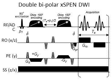
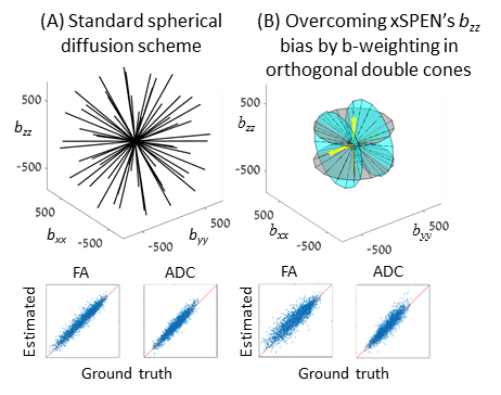
As in other SPEN based sequences [2,3], the sequential encoding and decoding occurring along xSPEN’s imaging axis results in a localized b-weighting which can be described by an extension of the Karlicek-Lowe diffusion formalism [4]:$$$\textbf{b}(t,r_0)=\gamma^2\int_{0}^{t}(\vec{\nabla}(\phi(t',r_0)\vec{\nabla}(\phi(t',r_0)^\text{T})dt'$$$, where $$$\phi(t',r_0)$$$ is the localized phase evolved by spins until time t’ under both the imaging and diffusion manipulations. A dominant contribution to b(t,ro) arises in xSPEN from the gradient acting along the slice-select axis, which is on throughout the scan. Gradient strengths in clinical scanners are insufficient for modulating further the diffusion weighting along this axis. Thus, an alternative gradient scheme was devised whereby the low-bandwidth dimension (y in Fig 1) is decoded twice, in two alternate sets of experiments that swap the readout (x) and slice-select (z) axes. As the slice-select gradient dominates the b-weighting this means that, for each of these acquisitions, the diffusion will be dominated by orthogonal bii directions. Bipolar diffusion modules applied along the remaining orthogonal orientations (see sequence in Figure 1) will further modulate the effective sampling of the b-space, leading to sets of conic shapes rotated about bii. The combination of two orthogonal xSPEN acquisitions then leads to a sufficiently extensive sampling of the full b-space, to enabling the retrieval of DTI parameters. Figure 2 illustrates this form of sampling the reciprocal diffusion space, and compares it to a standard spherical diffusion scheme.Methods
Sequence validation: To confirm the reliability of the double-cone approach, a set of 2601 sample tissues were simulated, with each tissue having a random proton density and a characteristic diffusion tensor with randomized directionality and eigenvalues spanning realistic fractional anisotropy (FA) and apparent-diffusion-coefficient (ADC) values. The signals arising from these different tissues under the action of the scheme in Fig. 2B were simulated, and additive Gaussian noise was added to the simulated signals so that the mean SNR for the b0 set would be 7%. Based on these synthetic sets, FA and ADC values were then estimated, and both mean absolute difference and r2 values were calculated against the ground truth FA and ADC values for various test gradient schemes.
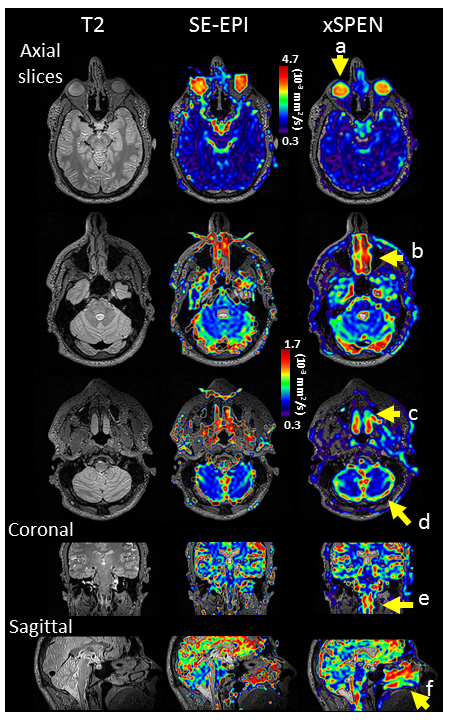
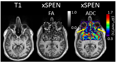
Scans: Human head images were acquired at 3T using a Siemens Trio scanner and a 32-channels head coil. A volunteer was scanned with the xSPEN DTI scheme at full brain coverage (4mm isotropic resolutions, b=1000 s/mm2, 4 repetitions), and with restricted FOV (3mm isotropic, b=800 s/mm2, 2 repetitions). EPI-DTI was run with the same b-values and a standard 32-direction scheme. For anatomical reference, T1- and T2-weighted scans were included.
Results
Figure 2B examines the new gradient scheme, by evaluating two planar DTI acquisitions performed in orthogonal orientations (gray and blue), each involving 15 bipolar diffusion modules whose directions were uniformly spread on the orthogonal plane. The scheme provided 0.89 and 0.91 r2 fit values for the FA and ADC respectively, vis-à-vis 0.95 and 0.96 values obtained by the standard scheme (Figure 2A and 2B, bottom). Figure 3 compares a 4mm isotropic xSPEN ADC map obtained with this scheme on a human head, against comparable results arising from SE-EPI. Notice how xSPEN preserves the anatomical features and provides diffusion information on regions challenged by strong inhomogeneity effects (Figure3, a-f) that are distorted or completely absent in SE-EPI. Figure 4 shows an additional example with a 3mm isotropic zoomed-in DTI parametric maps of the optic nerve region, where xSPEN preserves anatomical features and provides both FA and ADC values, despite the strong susceptibility effects by air interfaced to tissue and muscle in this region.Discussion and Conclusion
Thanks to its high resilience to field inhomogeneities, xSPEN DTI succeeds in characterizing key brain regions that are usually inaccessible by traditional single-shot methods. Diffusion information from those regions can open new opportunities in basic and clinical investigations.Acknowledgements
Funding by grants ISF 795/13, ERC-2014-PoC # 633888, Minerva Foundation #712277, and the Kimmel Institute of Magnetic Resonance (Weizmann) is acknowledged. We are also grateful to Dr. Sagit Shushan (Wolfson Medical Center), and the Weizmann MRI team (Edna Furman-Haran, Fanny Attar and Nachum Stern).References
[1] Zhang, Zhiyong, Amir Seginer, and Lucio Frydman. "Single-scan MRI with exceptional resilience to field heterogeneities." Magn Reson Med; doi:10.1002/mrm.26145 (2016).
[2] Shrot, Yoav, and Lucio Frydman. "The effects of molecular diffusion in ultrafast two-dimensional nuclear magnetic resonance." The Journal of chemical physics 128.16 (2008): 164513.
[3] Solomon, Eddy, Noam Shemesh, and Lucio Frydman. "Diffusion weighted MRI by spatiotemporal encoding: analytical description and in vivo validations." Journal of Magnetic Resonance 232 (2013): 76-86.
[4] Karlicek, R. F., and I. J. Lowe. "A modified pulsed gradient technique for measuring diffusion in the presence of large background gradients." Journal of Magnetic Resonance (1969) 37.1 (1980): 75-91.
Figures