5035
A feasibility study of MR T2-mapping for evaluating birth-related levator ani muscle injury1Department of Radiology, Tianjin First Center Hospital, Tianjin, People's Republic of China, 2Philips Healthcare, Beijing, China, beijing
Synopsis
Currently, the abnormal morphology of LAM is assessed with an unaided subjective diagnosis. But T2 mapping can quantitatively evaluate the injury muscle and indicate the injured degree and region of the LAM. In this study, we investigate whether the quantitative MR T2-mapping can evaluate birth-related levator ani muscle injury. The results showed that T2-mapping can quantitatively assess birth-related levator ani muscle injury and T2-mapping color-coded images show the range and degree of LAM injury visually. Moreover, it is helpful for detecting micro lesions which is difficult for just using PDWI images.
Purpose
As we all know, vaginal delivery often accompany with levator ani muscle(LAM) injury, which is the important support structure for holding the pelvic organs to normal position. The early detection and treatment of injured LAM is most urgent for clinic. Currently, the abnormal morphology of LAM is assessed with an unaided subjective diagnosis, and T2 mapping can quantitatively evaluate the injury muscle and indicate the injured degree and region of the LAM. The aim of this study is to investigate whether the quantitative MR T2-mapping can evaluate birth-related levator ani muscle injury.
Materials and methods
Twenty-five primiparas about six weeks after first vaginal delivery and twelve nulliparous women were included in this study and divided into primiparous and control groups. All the subjects underwent pelvic MRI examinations including T2-mapping and PDWI imaging with a 3T MRI scanner (Ingenia, Philips Healthcare, the Netherlands). Primiparous group was furtherly divided into two subgroups: levator ani muscle (LAM ) subdivision(Puborectal muscle, PRM; Iliococcygeal muscle, ICM)injury group and non-injury group according to whether there were edema, avulsion, or rupture in LAM subdivision based on PDWI images. Two radiologists with more 3 years diagnostic experience evaluated the T2 values of PRM, ICM and observed T2-mapping color-coded images respectively. The consistency between two observers for T2 values of PRM, ICM were evaluated using the intraclass correlation coefficient (ICC). The difference of T2 values in each LAM subdivsion among control group, non-injury group and LAM injury group were analyzed using ANOVA and followed by post-hoc multiple comparison with Scheffe method.Results
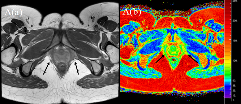
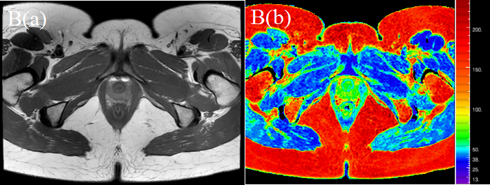
There were thirteen PRM injury cases and twelve non-injury cases in primiparous group according to PDWI images, and we did not find ICM injury cases in our study. Inter-rater reliability for T2 values calculated by two observers were good (ICC>0.75). The T2 value of PRM in PRM injury group was the highest among three groups, and T2 values in PRM injury group, non-injury group and control group were 62.78±1.23ms, 49.75±3.17ms and 49.96±4.37ms respectively. There were significant differences between PRM injury group and non-injury group (P<0.001), as well as between PRM injury group and control group (P<0.001). And there was no significant difference between non-injury group and control group (P=0.989). The T2 values of ICM in PRM injury group, non-injury group and control group were 70.80±6.50ms, 62.41±7.32ms and 62.78±6.91ms respectively. And there were significant differences between PRM injury group and non-injury group(P<0.001), as well as between PRM injury group and control group(P<0.001). Moreover, there was no significant difference between non-injury group and control group(P=0.981). T2 mapping color-coded images showed PRM color gradation in PRM injury group were mixed with blue, green, and yellow, and the tone were bright(Fig.1). ICM color gradation were uneven with green and yellow, and the tone were brighter than those of control group and non-injury group(Fig.2).Discussion
The increase of T2 value indicated that the tissues suffer from some edema and injury[1-2]. In the PRM injury group, T2 value of PRM is higher than that of non-injury group, which is consistent with physiological conditions of injured muscle, indicating that T2 mapping can reflect injured muscle situation. And the T2 value of ICM in the PRM injury group is also higher than that of non-injury group, which may indicate that some potential injury existed in ICM of PRM injury group.Conclusions
T2-mapping can quantitatively assess birth-related levator ani muscle injury and T2-mapping color-coded images show the range and degree of LAM injury visually. Moreover, T2-mapping technique is more effective in the detection of micro lesions than PDWI imaging.Acknowledgements
No acknowledgement found.References
Reference:
1.Ha DH, Choi S, Kang EJ, et al. Diffusion tensor imaging and T2 mapping in early denervated skeletal muscle in rats[J]. J Magn Reson Imaging, 2015, 42(3): spcone.
2.Yan D, Zhang J, Liang W, et al. Magnetic resonance imaging and histopathological analysis of experimental muscle injuries in a rabbit[J]. Biomed Environ Sci, 2013, 26(10): 841-848.
Figures