5034
Utility of Radial Reformation of Three-dimensional Fat-suppressed Multi-echo Gradient-recalled-echo Imaging in the Evaluation of Acetabular Labral Injuries and Femoroacetabular Impingement1Chugoku Rosai Hospital, Kure, Japan, 2Hiroshima Prefectural Hospital, Hiroshima, Japan, 3Hiroshima University Hospital, Hiroshima, Japan
Synopsis
Radial images through the center of the acetabulum in the plane including the entire acetabular rim are useful for evaluating acetabular labral injuries, and those perpendicular to the center of the femoral head through the central axis of the femoral neck enable evaluation of the morphological findings of femoroacetabular impingement. Continuous thin-slice images from optimized 3D fat-suppressed multi-echo gradient-recalled-echo allow 360° radial reformation of hip joints, offering precise evaluation of the range of acetabular labral injury and femoroacetabular impingement morphology.
PURPOSE
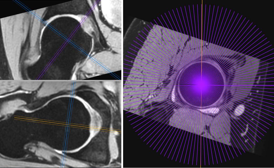
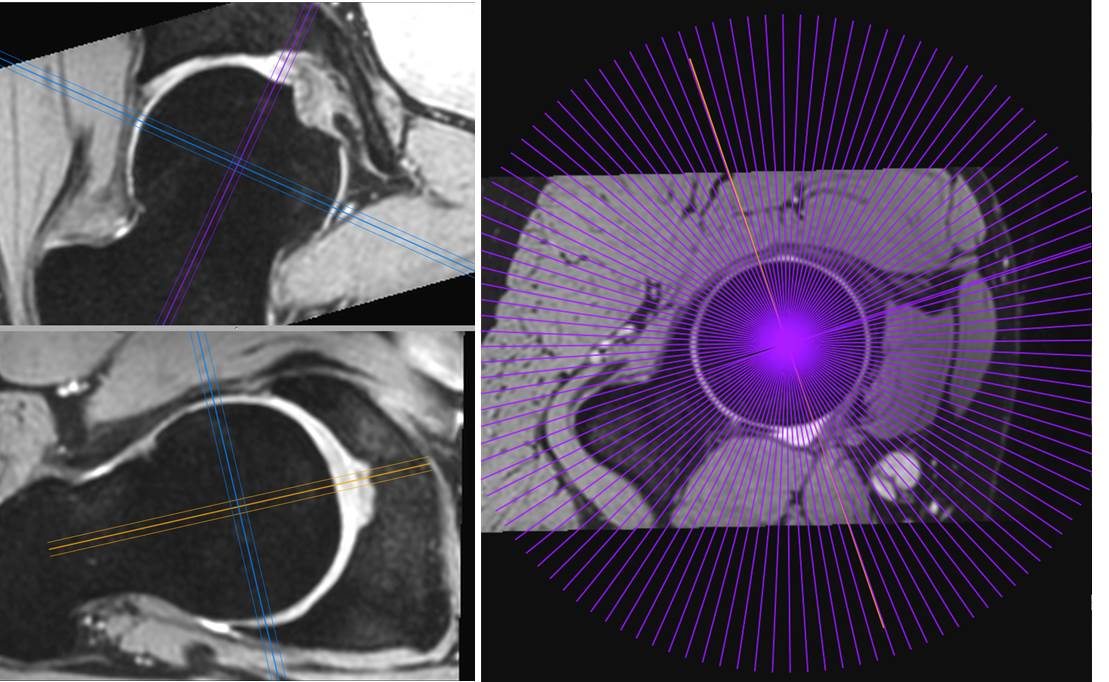
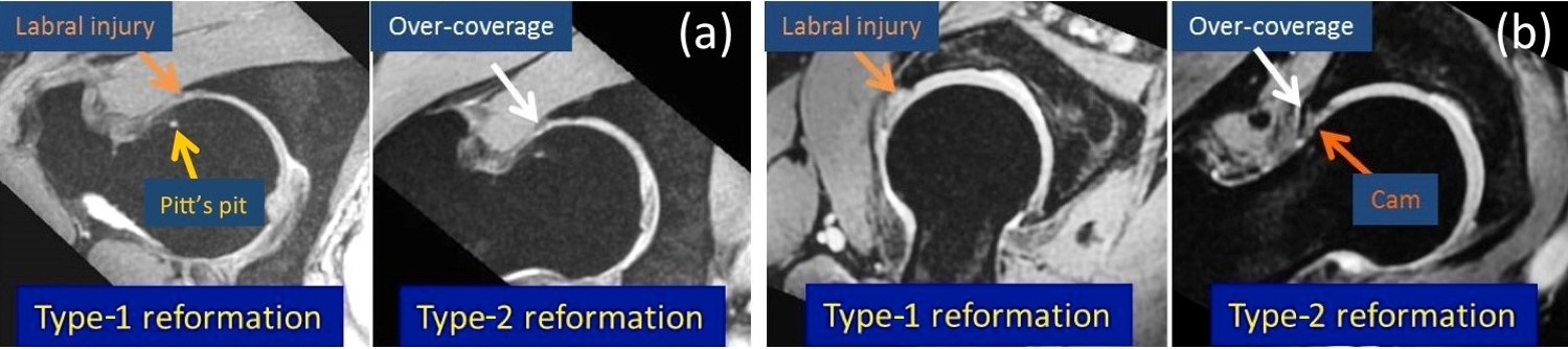
The evaluation of acetabular labral injuries and femoroacetabular impingement (FAI) is difficult using standard two-dimensional (2D) axial, coronal, and sagittal magnetic resonance imaging (MRI).1,2 Advantages of three-dimensional (3D) isotropic acquisitions of MRI include a high signal-to-noise ratio, high spatial resolution, and the ability to yield high-quality multiplanar images from the original acquisition dataset. The first purpose of this study was to evaluate the diagnostic accuracy of radial reformation through the center of the acetabulum perpendicular to the plane across the entire acetabular rim (type-1 radial reformation; Fig. 1) in 3D fat-suppressed multi-echo gradient-recalled-echo (FS multi-echo GRE) imaging for evaluating acetabular labral injury by comparison with 2D radial T2*-weighted imaging (T2*WI).3 The second purpose was to confirm the utility of radial reformation perpendicular to the central axis of the femoral head and neck (type-2 radial reformation; Fig. 2) in 3D FS multi-echo GRE imaging for evaluating FAI in patients with labral injury.METHODS
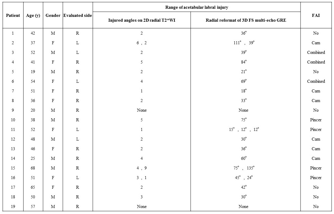
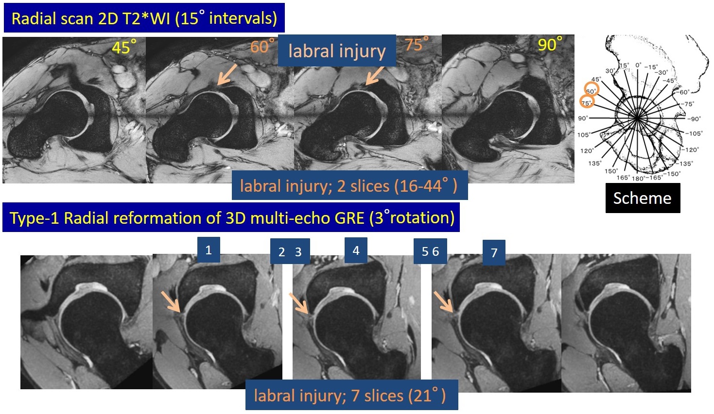
We examined 19 patients (mean age, 44.2 ± 13.6 years; 10 men, 9 women) with suspected acetabular labral injury using a Philips Ingenia 3.0-T system. We optimized axial 3D FS multi-echo GRE imaging for evaluating acetabular labral injury (TR, 32 ms; TE, 2.3, 5.6, 8.9, 12.2, and 15.5 ms; flip angle, 7°; slice thickness, 0.5 mm; pixel size, 0.5 ´ 0.5 mm). We also acquired 2-mm slice thickness type-1 and type-2 radial reformations at 3° intervals of 3D FS multi-echo GRE imaging of the right diseased hip joints of 13 patients and the left diseased hip joints of 6 patients using OsiriX on Mac OS X. In addition, we obtained 12 radial slices at 15° intervals of 2D T2*WI through the center of the acetabulum perpendicular to the plane across the entire acetabular rim (TR, 400 ms; TE, 18.4 ms; flip angle, 30°; slice thickness, 4 mm; pixel size, 0.5 ´ 0.5 mm). The range of acetabular labral injury of each patient was evaluated by the consensus decision of two experienced radiologists. Two weeks after interpreting 2D T2*WI, the same radiologists evaluated labral injury on type-1 radial reformation of 3D FS multi-echo GRE imaging. Diagnostic accuracy using the type-1 radial reformation was assessed in comparison with 2D radial T2*WI. The same radiologists estimated the degree of FAI using type-2 radial reformation of 3D FS multi-echo GRE imaging. An α angle over 55° was considered indicative of cam-type FAI.4 Pincer-type FAI was diagnosed based on over-coverage of the acetabulum.2RESULTS
We identified 2 patients with no labral injury, and labral injury in the anterosuperior zone of acetabula in 10 patients, posterosuperior zone in 3 patients, posteroinferior zone in 1 patient, and both anterosuperior and posterosuperior zones in 3 patients from type-1 radial reformation of 3D FS multi-echo GRE imaging and 2D radial T2*WI. The rate of diagnostic concordance was 100% (19/19). In each patient with acetabular labral injury, the injured range demonstrated on type-1 radial reformation of 3D FS multi-echo GRE imaging included the injured angles on 2D radial T2*WI (Table 1, Fig. 3). Type-1 radial reformation of 3D FS multi-echo GRE imaging revealed two small labral injuries not detected on 2D radial T2*WI in 1 patient. We could evaluate FAI in all patients (no FAI, n=6; cam-type, n=6; pincer-type, n=4; combined cam- and pincer-type, n=3) using type-2 radial reformation of 3D FS multi-echo GRE imaging (Table 1, Fig. 4).
.
DISCUSSION
Due to reduced partial volume averaging by acquiring thin continuous slices, 3D pulse sequences are advantageous in evaluating abnormalities of the ball-and-socket-shaped hip joint, and 3D multi-echo GRE imaging yields higher signal-intensity images than conventional 2D T2*WI with single echo acquisition. Radial reformation at small intervals of 3D FS multi-echo GRE imaging shows the precise range of acetabular labral injury in comparison with 2D radial T2*WI. Moreover, radial reformation perpendicular to the central axis of the femoral head and neck permits a better appreciation of hip joint contours than X-ray films, and allows evaluation of cam- and pincer-type FAI. Potential disadvantages of 3D GRE imaging include relatively long acquisition times and inherent sensitivity to susceptibility artifacts, which may be problematic in postoperative imaging.5 One weakness of this study was the lack comparison with findings from MRI and arthroscopy.CONCLUSION
Type-1 and -2 radial reformation of 3D FS multi-echo GRE imaging were useful for evaluating the range of acetabular labral injury and determining whether patients with labral injury have FAI, respectively.Acknowledgements
No acknowledgement found.References
1. Park SY, Park JS, Jin W, et al. Diagnosis of acetabular labral tears: comparison of three-dimensional intermediate-weighted fast spin-echo MR arthrography with two-dimensional MR arthrography at 3.0 T. Acta Radiologica. 2013;54(1):75-82.
2. Pfirrmann CW, Mengiardi B, Dora C, et al. Cam and pincer femoroacetabular impingement: characteristic MR arthrographic findings in 50 patients. Radiology. 2006;240(3):778-785.
3. Horii M, Kubo T, Hirasawa Y. Radial MRI of the hip with moderate osteoarthritis. Bone & Joint Journal. 2000;82(3):364-368.
4. Nouh MR, Schweitzer ME, Rybak L, et al. Femoroacetabular impingement: can the alpha angle be estimated? AJR Am J Roentgenol. 2008;190(5):1260-1262.
5. Naraghi A, White LM. Three-dimensional MRI of the musculoskeletal system. AJR Am J Roentgenol. 2012;199(3):W283-W293.
Figures