5033
Clinical interpretation of asymptomatic medial collateral ligament injury observed on magnetic resonance imaging in adolescent baseball players1University of Tsukuba, Tsukuba, Japan
Synopsis
This presentation clarified the clinical interpretation of 'asymptomatic MCL injury of the elbow observed on MRI among adolescent baseball players' by comparing the findings of MRI with those of standard clinical orthopedic examinations including palpation and US. Our results suggested the finding includes a broad spectrum of injuries; namely, a group of injuries that are characterized by changes associated with 'adaptation' and group of a 'pre-injury' characterized by changes to symptomatic MCL injury.
Purpose
In 2014, we started applying low-field (0.18 T) MRI of the extremities for the screening of adolescent baseball players with no clinical symptoms. With this method, all (100%) 63 subjects aged 9 to 12 years completed the examination, and we acquired MRI images of sufficient quality for diagnosis1. As the MRI machine was of the open-type, the subjects could talk to their parents and examiners, which helped them to relax. Low-field MRI may be useful for the screening of elbow injuries in adolescent baseball players. This preliminary results showed that there were no cases of osteochondritis dissecans or avulsion fractures of the medial epicondyle among the study subjects, which are both frequently encountered in the daily practice by orthopedicians. However, the frequency of injury of the medial collateral ligament (MCL) itself, which does not frequently occur in adolescent players, was unexpectedly high at 41.9% (26/63) 1. We wondered why the subjects showed no clinical symptoms despite evidence of MCL injury on MRI. The purpose of this study was to clarify the clinical interpretation of this ‘asymptomatic MCL injury of the elbow observed on MRI among adolescent baseball players’ by comparing the findings of MRI with those of standard clinical orthopedic examinations including US and palpation.
Methods
Sixty-four male and female baseball players (mean age, 11.2 [range, 9–13] years) were enrolled. MRI was performed by an open-type 0.18 T MRI C-SCAN (Esaote Inc., Ferrara, Italy) (Figure 1). MCL function was evaluated by measuring the opening of the ulnohumeral joint width at the level of the anterior band using US (Figure 2). Palpation included MCL tenderness, the moving valgus test, and the milking test. The correlation between MRI and ultrasonography findings (t-test), and MRI and palpatioin ( χ2 test and Fisher exact test ) were analyzed.Results
Thirty-four subjects (53.1%) showed ‘asymptomatic MCL injury’ findings (Figure 3) in this study. The mean laterality of the cleft between articulations showed no significant difference (P = 0.16) between the group with (0.29 ± 1.06 mm) and without (0.08 ± 0.96 mm) MCL injury on MRI. There was no correlation between subjects with MCL tenderness and MCL injury on MRI and positive moving valgus test and MCL injury on MRI. However, there was a weak correlation between subjects with positive Milking test and MCL injury on MRI (φ coefficient = 0.3, P = 0.02).Discussion
In the present study, there were no statistically significant differences in ligament function assessed by US. This means that most adolescent baseball players who have findings of ‘asymptomatic MCL injury of the elbow on MRI’ also have normal ligament function. Asymptomatic MCL injury of the elbow might be caused by overuse, which is a slight but repetitive type of injury. Therefore, it is possible that subjects show no apparent clinical symptoms and normal ligament function because the extent of each single stress is very minor. However, although each single stress is very minor, repetitive stimulation can cause structural changes in the MCL. In this situation, subjects might not notice this gradual damage to their elbow, and thus, remain asymptomatic for a long period of time. For these reasons, we consider that MRI findings of MCL injury of the elbow can be present despite no apparent clinical symptoms and normal ligament function in active adolescent baseball players.
This situation is very similar to the characteristics of “adaptation,” which have been reported in many previous studies2, 3. “Adaptation” refers to normal MCL thickening on MRI in baseball players showing normal ligament function and no apparent clinical symptoms. This is observed in players after epiphyseal closing around the elbow. Although, adaptation can been considered a normal MCL change, the mechanism of ligament thickening is unclear. We assumed that in adolescent baseball players with asymptomatic MCL injury of the elbow diagnosed by MRI, adaptation might represent chronic elbow injury after the cure of an overuse injury. Therefore, we hypothesized that asymptomatic MCL injury diagnosed by MRI in adolescent baseball players might represent the transition period in which adaptation occurs. Moreover, there was weak correlation between positive Milking test and positive MCL injury on MRI in this study. Asymptomatic subjects might therefore fall into the “symptomatic” MCL injury group (a pre-injury group).
Conclusion
We consider that asymptomatic MCL injury diagnosed by MRI in adolescent baseball players includes a broad spectrum of injuries; namely, a group of injuries that are characterized by changes associated with adaptation and group of a pre-injury characterized by changes to symptomatic MCL injury (figure 4).Acknowledgements
I would like to express my gratitude to Shunpei Miyakawa for permission to use extrimity dedicated MRI machine.References
1. Okamoto Y, Maehara K, Kanahori T, et al. Incidence of elbow injuries in adolescent baseball players: screening by a low field magnetic resonance imaging system specialized for small joints. Jpn J Radiol. 2016;34(4):300-306.
2. Marshall NE, Keller RA, Van Holsbeeck M, et al. Ulnar Collateral Ligament and Elbow Adaptations in High School Baseball Pitchers. Sports Health. 2015;7(6):484-488.
3. Ellenbecker TS, Mattalino AJ, Elam EA, et al. Medial elbow joint laxity in professional baseball pitchers. A bilateral comparison using stress radiography. Am J Sports Med. 1998;26(3):420-424.
Figures
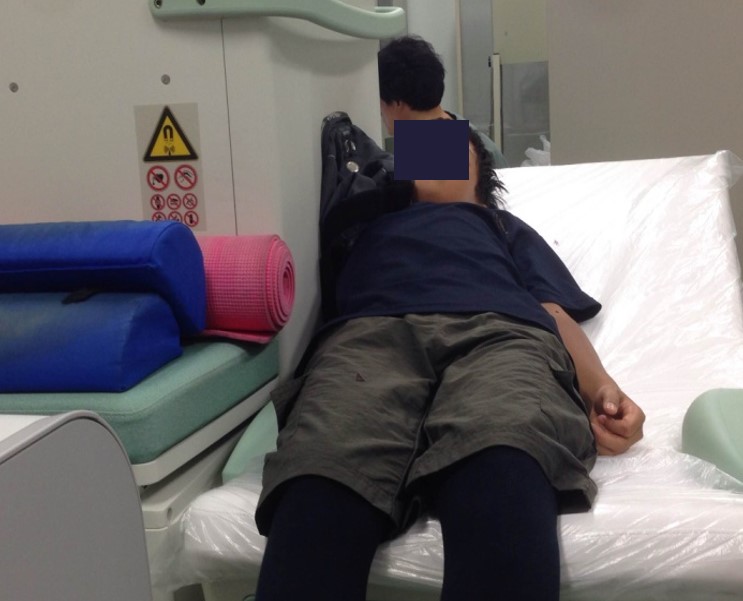
Figure 1
An 12-year-old girl having an MRI check-up for elbow injuries induced by baseball using a 0.18 T low magnetic field machine. Her body is positioned mainly on the bed and her right arm is inserted into the MRI machine.
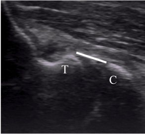
Figure 2
The white line represents the most distal point of the medial trochlea and measurement of the vertical distance across the joint to the ulnar coronoid using the 1-line method. Abbreviations: C, coronoid process; T, trochlea
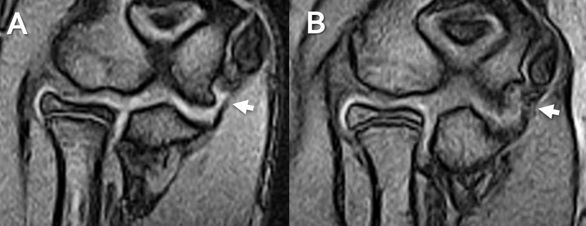
Figure 3
A; The subject was a 13-year old pitcher. Intact MRI of the MCL (arrow) B; The subject was a 12-year-old pitcher. Diffuse swelling and elevation of signal intensity were observed (arrow).
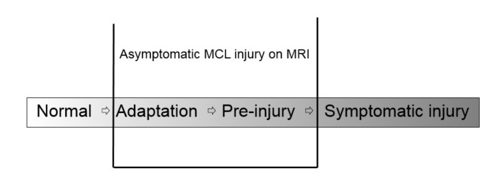
Figure 4
From the characteristics of injury of adolescent baseball players, the literature on ligament injury, and the concept of adaptation, we hypothesize that asymptomatic MCL injury includes a broad spectrum of injuries. In particular, it would be the ‘adaptation’ group in the near future, and the 'pre-injury' group in the future.