5025
Age-related differences in quantitative muscular contraction parameters measured by synchronous dynamic MRI of electrical muscle stimulation1Radiology, Division of Radiological Physics, University of Basel Hospital, Basel, Switzerland, 2Biomedical Engineering, University of Basel, Basel, Switzerland, 3Sports Medicine, University of Basel, Basel, Switzerland, 4Musculoskeletal Radiology, Clinic of Radiology and Nuclear Medicine, University of Basel Hospital, Basel, Switzerland, 5Ricerche Diagnostiche Srl, Bari, Italy
Synopsis
Stimulation of the quadriceps muscle group of the thigh and synchronous phase contrast imaging at a 3T MRI scanner were applied to six young and 13 elderly volunteers, to assess age-related differences. Dynamic strain and strain rate maps were reconstructed. Age-related differences both for strain and strain rate were observed and the significance of the difference increased when the vastus intermedius muscle was included.
INTRODUCTION
As it has been suggested that age induces changes in the contractility and elastic properties of the skeletal muscle1, 2, the purpose of this study was to investigate whether strain and strain rate, assessed by synchronizing the MRI acquisition with a commercial electrical muscle stimulator (EMS)3, differ between young and elderly volunteers; serving as a baseline for future studies on muscle disease.METHODS
For the experimental setup, a commercially-available EMS device was used to induce involuntary periodic muscle contraction of the vastus lateralis muscle (VL), synchronized with high-temporal-resolution cine phase contrast (PC) MRI acquisition at 3T3-5. A three-directional gradient echo PC velocity encoding sequence was applied. MR acquisitions were performed on a parasagittal slice with a spatial resolution of 2.3x2.3x5 mm3 and a temporal resolution of 42 ms. The velocity encoding had a VENC of 25 cm/s (TR/ TE = 10.6/ 7.21 ms, bandwidth/ pixel = 400 Hz / Px, flip angle = 10°, FOV= 225 x 300 mm2, 1 k-space line per segment, acquisition time 5 min) and 94 temporal phases were acquired. Each contraction lasted 1 sec (i.e., 1 sec ramp time, 1 sec plateau, 1 sec ramp down, 2 sec relaxation)3. Strain rate (SR) and strain (S) vectors were extracted from the velocity fields3, 1, 4.
Two groups of healthy volunteers were included in the study: a) six male younger volunteers with an average age of 29 ± 6 years (21-35y), b) 13 older volunteers (6 female, 7 male) with an average age of 74 ± 3 years (70-82y).
The method was applied to the younger healthy volunteers with a fixed stimulation current of 18 mA to stimulate the VL muscle of the thigh. For the elderly volunteers, the stimulation was applied twice, once at 18 mA for direct comparison with the younger group, and the second at a higher level of 22mA. The post-processing analysis was performed twice: a) for a region-of-interest (ROI) covering the VL only, and b) for a broader ROI comprising also the vastus intermedius (VI) muscle. Larger ROIs were also included assuming that the contractile properties are more dependent on the fiber type than on the muscle6.
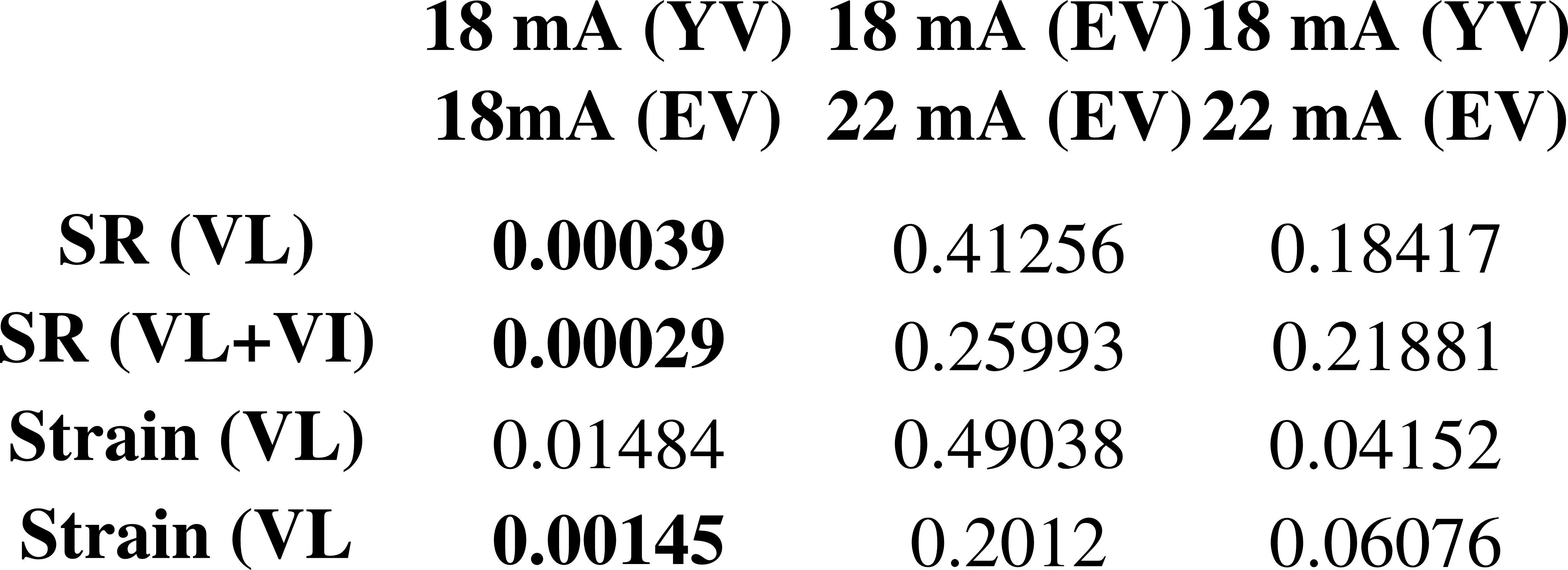
Finally, two-sample unpaired t-test was performed to assess the difference in strain and strain-rate magnitude values. Since 12 separate comparisons were performed (i.e., S and SR on VL and VL+VI ROIS for 18mA and 22mA stimulations in elderly and 18mA in younger volunteers), a Bonferroni correction for multiple parameter comparisons was applied, thus lowering the significance level to p = 0.004.
RESULTS
In general, the elderly volunteers were less sensitive to the stimulation current. In most of the cases (10/13), it was necessary to use 22 mA in the elderly to visually achieve the same displacements as of 18 mA in the younger volunteers.
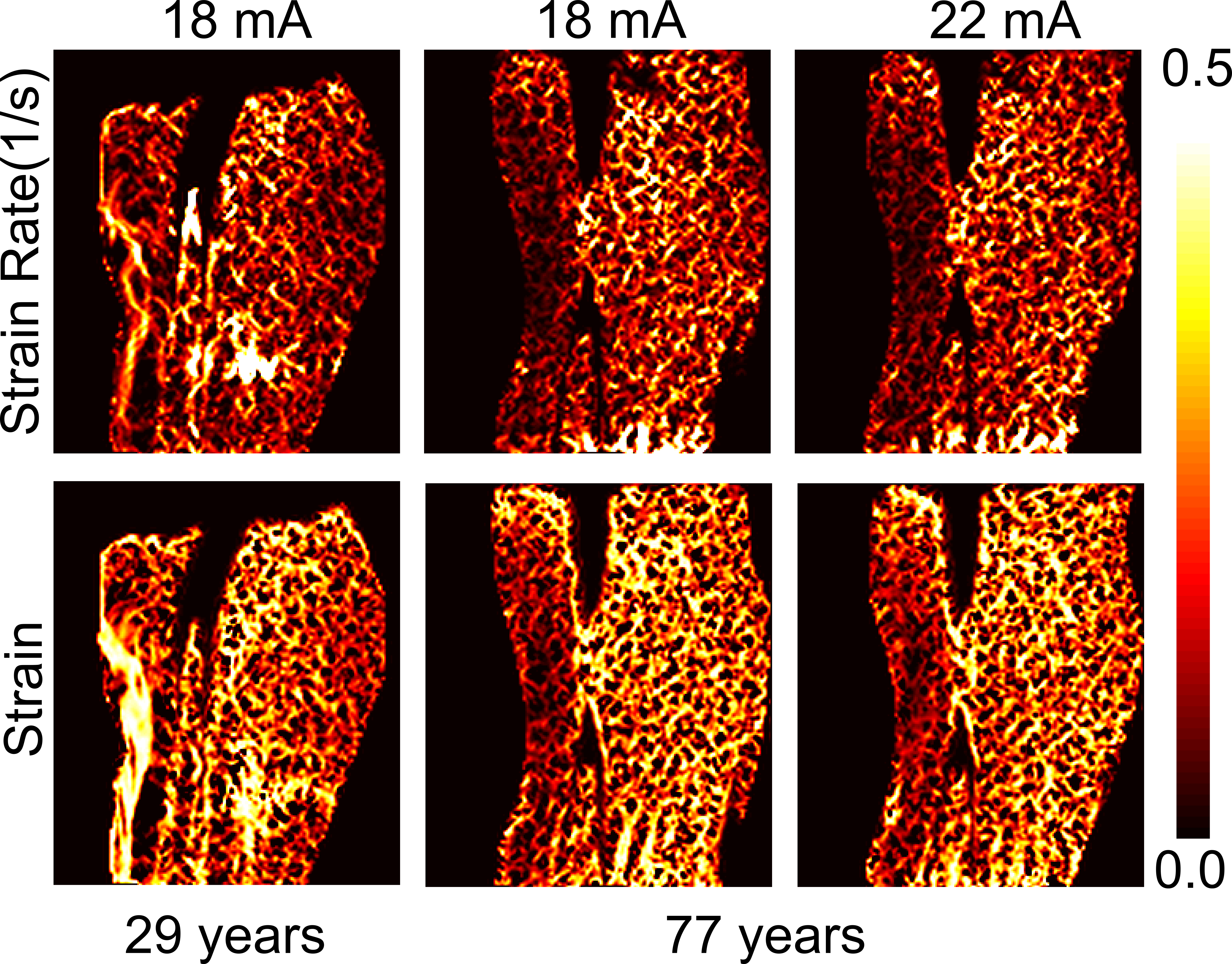
A qualitative evaluation of strain and strain rate maps revealed a clearly defined region with increasing values during contraction in most of the young volunteers (see Fig. 1). Conversely, in the elderly volunteers, the strain and strain rate values were lower and mostly without a clearly activated region in the stimulated muscle.
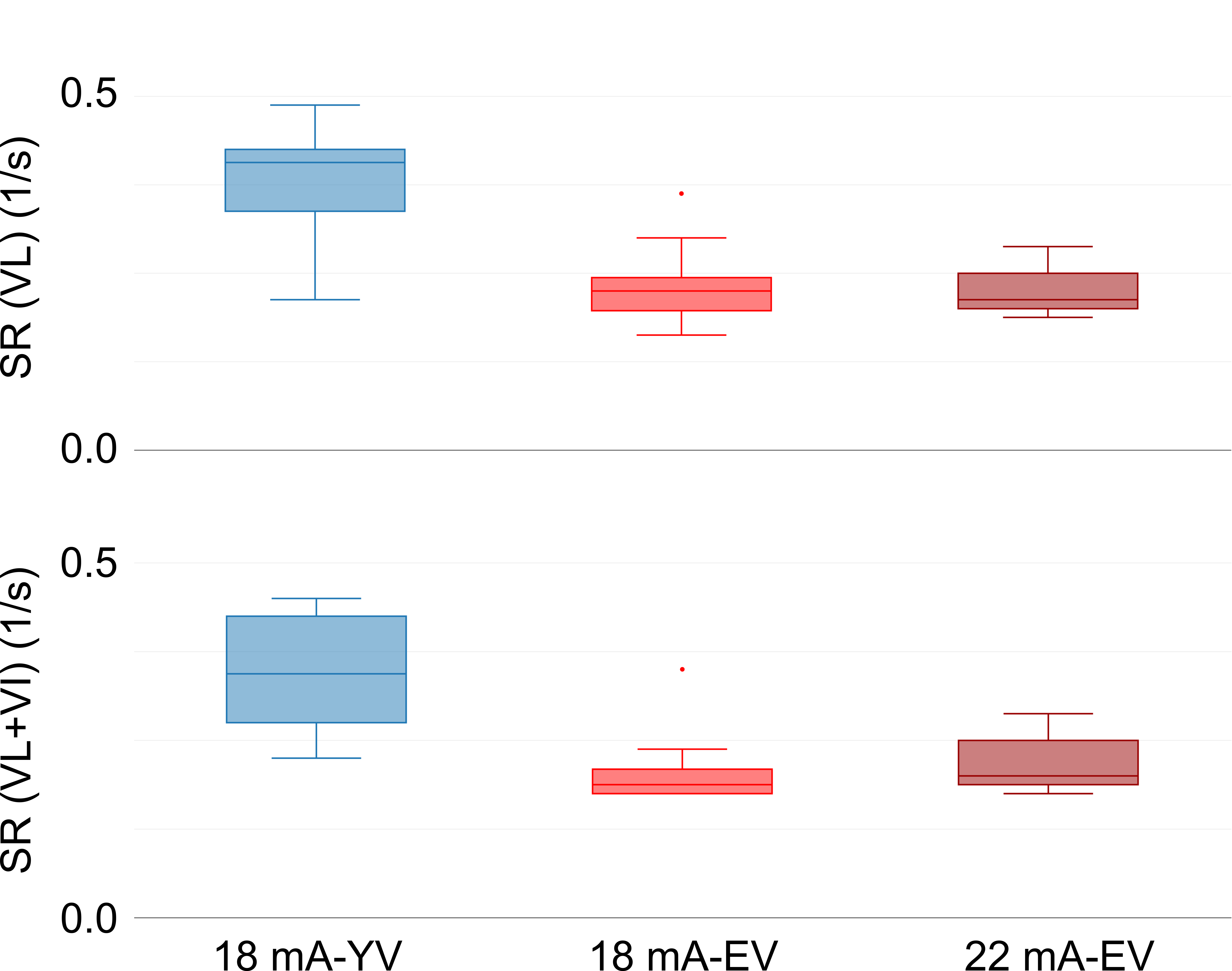
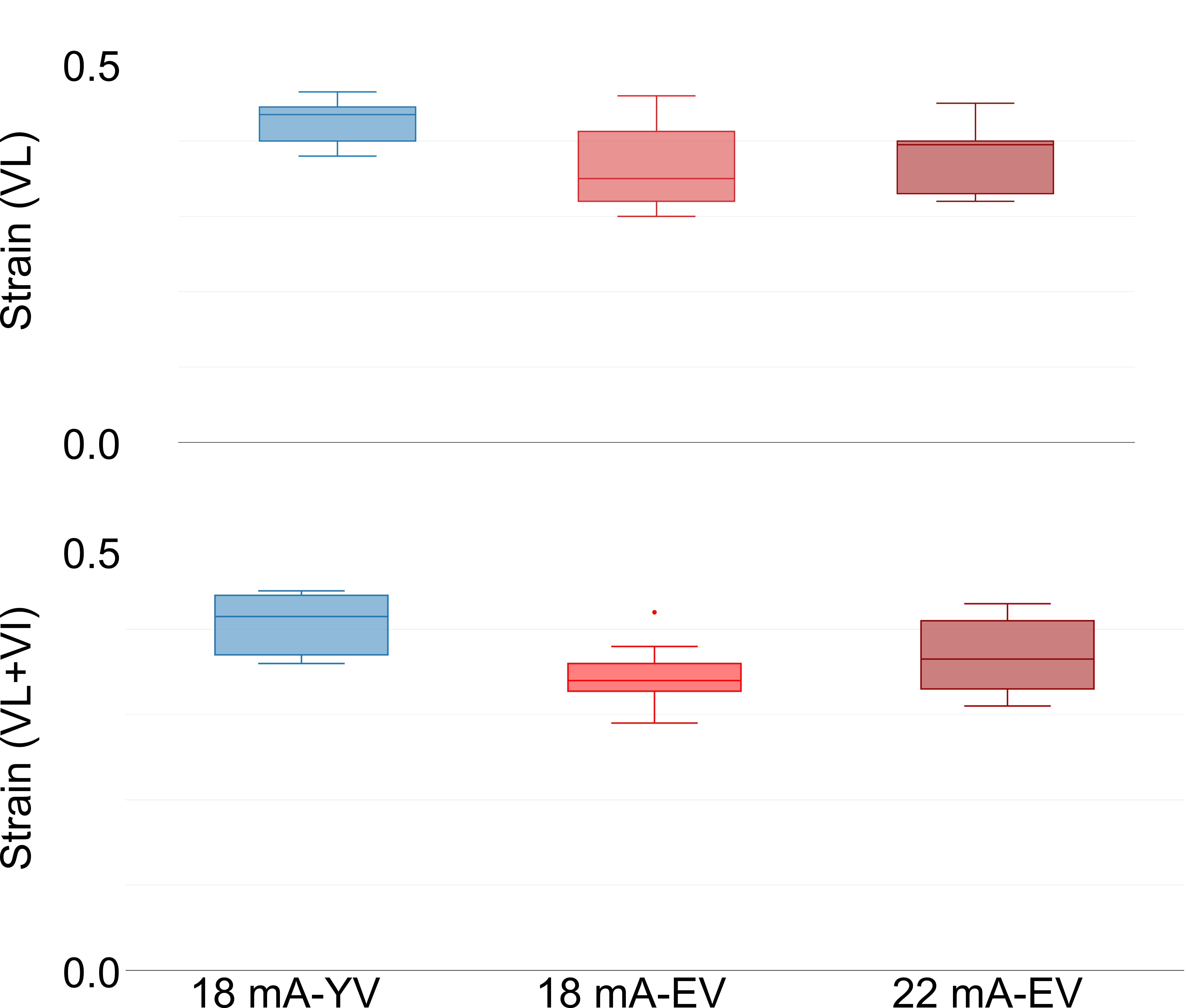
The quantitative evaluation showed that a stimulation of 18 mA on younger volunteers yielded higher strain rate values than 18 or 22 mA on elderly volunteers and that the difference between the two groups was larger if the VI was included (see Fig. 2). Furthermore, strain values were higher for the younger volunteers and the inclusion of the VI increased this difference between the two groups as well.
Finally, comparing the differences regarding the measurements at 18 mA of young volunteers (YV) and elderly volunteers (EV) the t-tests revealed a significant difference for SR (VL), SR(VL+VI) and Strain (VL+VI), but not for SR(VL) (see Table 1). No significant differences were found between: a)18 mA YV and 22 EV, and b) 18 mA EV and 22 mA EV.
DISCUSSION
This study showed a significant impact of age on the functionality of the skeletal muscle contraction assessed non-invasively by the strain and strain rate parameters, even among healthy subjects. Moreover, the data indicates that the difference of strain rate and strain values between young and elderly volunteers increased when a broader ROI including both VL and VI was considered. One possible explanation for that could be preferential loss of type II fibers with age7.CONCLUSION
Strain and strain rate measurements with MRI of EMS-controlled involuntary muscle contraction show age-related differences and can therefore provide useful information about the physical condition of muscles.Acknowledgements
This work was supported by the Swiss Foundation for Research on Muscle Diseases (FSRMM).References
1. Sinha U., Malis V., Csapo R., Moghadasi A., Kinugasa R., and Sinha S. Age-Related Differences in Strain Rate Tensor of the Medial Gastrocnemius Muscle During Passive Plantarflexion and Active Isometric Contraction Using Velocity Encoded MR Imaging: Potential Index of Lateral Force Transmission, Magn Reson Med 2015; 73:1852–1863.
2. Ochala J., Frontera W., Dorer DJ., Van Hoecke J., Krivickas LS. Single skeletal muscle fiber elastic and contractile characteristics in young and older men, J Gerontol A Biol Sci Med Sci. 2007 Apr;62(4):375-81.
3. Deligianni X, Pansini M, Garcia M, Hirschmann A, Schmidt-Trucksäss A, Bieri O, Santini F. Synchronous MRI of muscle motion induced by electrical stimulation. Magn. Reson. Med. 2016. doi: 10.1002/mrm.26154.
4. Sinha S, Hodgson JA, Finni T, et al. Muscle kinematics during isometric contraction: development of phase contrast and spin tag techniques to study healthy and atrophied muscles. J Magn Reson Imaging 2004;20:1008–1019.
5. Drace JE, Pelc NJ. Measurement of skeletal muscle motion in vivo with phase-contrast MR imaging. J Magn Reson Imaging 1994;4:157–163.
6. Harridge SD, Bottinelli R, Canepari M, Pellegrino MA, Reggiani C, Esbjörnsson M, Saltin B. Whole-muscle and single-fibre contractile properties and myosin heavy chain isoforms in humans. Pflüg. Arch. Eur. J. Physiol. 1996;432:913–920.
7. Rogers MA, Evans WJ., Changes in skeletal muscle with aging: effects of exercise training, Exerc Sport Sci Rev. 1993;21:65-102.
Figures