5024
Simultaneous multi-slice TSE imaging of the hip joint: Acquisition time reduction and imaging quality.1radiology, Saitama Medical University Hospital, Saitama, Japan, 2orthopedics, Saitama Medical University Hospital, Saitama, Japan, 3Siemens Healthcare K.K, Tokyo, Japan, 4Siemens Healthcare GmbH, Erlangen, Germany, 5Siemens Healthcare, Minneapolis, United States
Synopsis
Simultaneous-Multi-Slice Turbo-Spin-Echo (SMS TSE) protocols were added to our conventional (CNV) hip joint MR examination in 21 patients. Image quality of acetabular labrum, articular cartilage, round ligament and bony trabeculae were evaluated by three reviewers and showed comparable image quality, whereas SMS was slightly better for bony trabeculae. If CNV is substituted by SMS, total scan time can be reduced by approximately 40%. Although SMS protocols showed higher specific absorption rate (SAR), it only accounted for 20% of scans which exceeded the first level of SAR limit. SMS could be applied to clinical examination with advantage of acquisition time reduction.
PURPOSE
MR imaging to delineate the fine components of the hip joint requires superb imaging quality with high spatial resolution. However, this tends to prolong the acquisition time. The SMS technique using multiband excitation pulse enables substantial reduction of the time required according to the number of slices. When SMS is utilized for scan time reduction with the TSE sequence, which is most widely used for joint imaging, scan parameters related to image contrast (e.g.TR) are changed. Moreover, SAR is increased. The purpose of this study was to prospectively compare the hip joint TSE imaging with and without SMS regarding acquisition time, imaging quality and SAR.METHODS
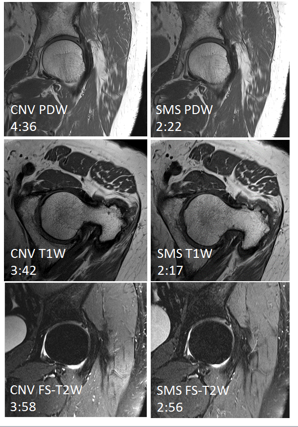
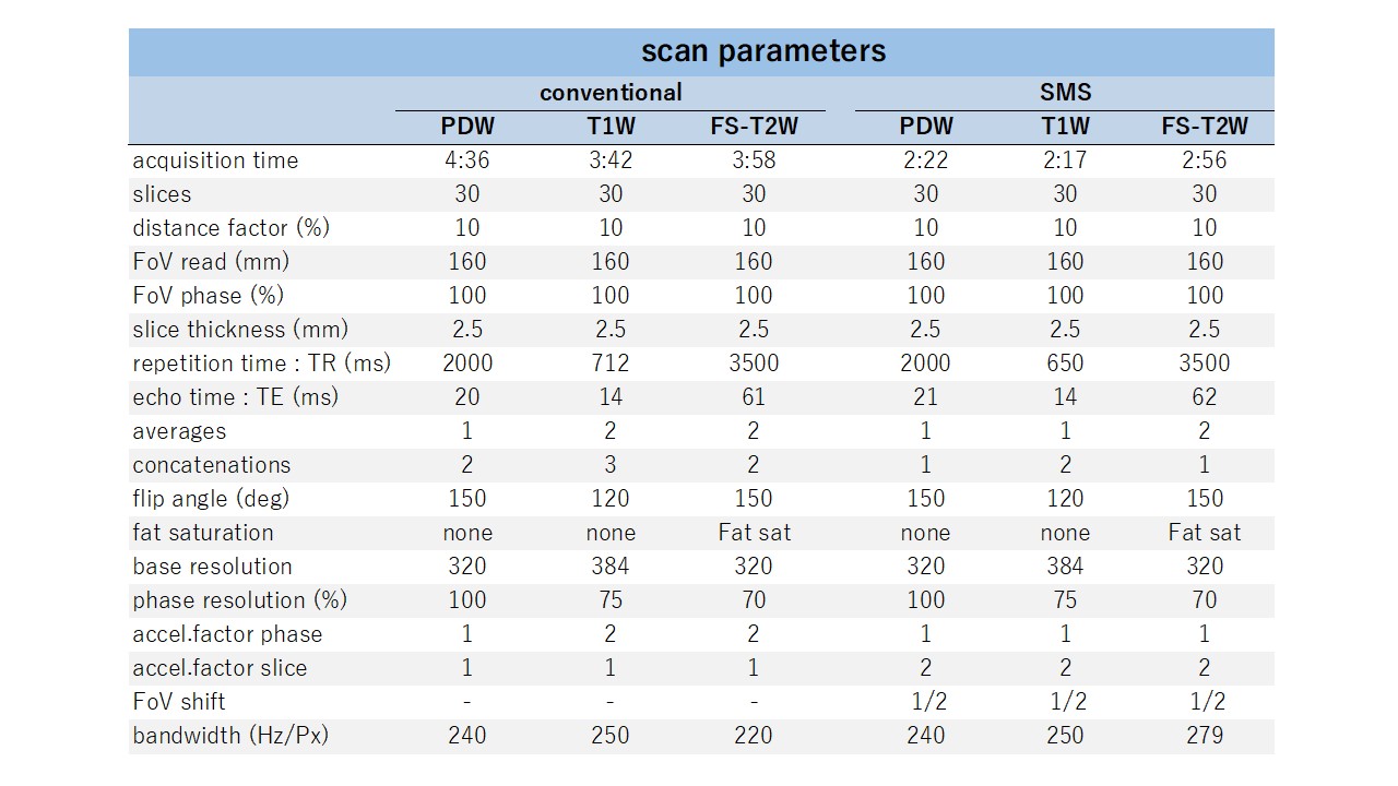
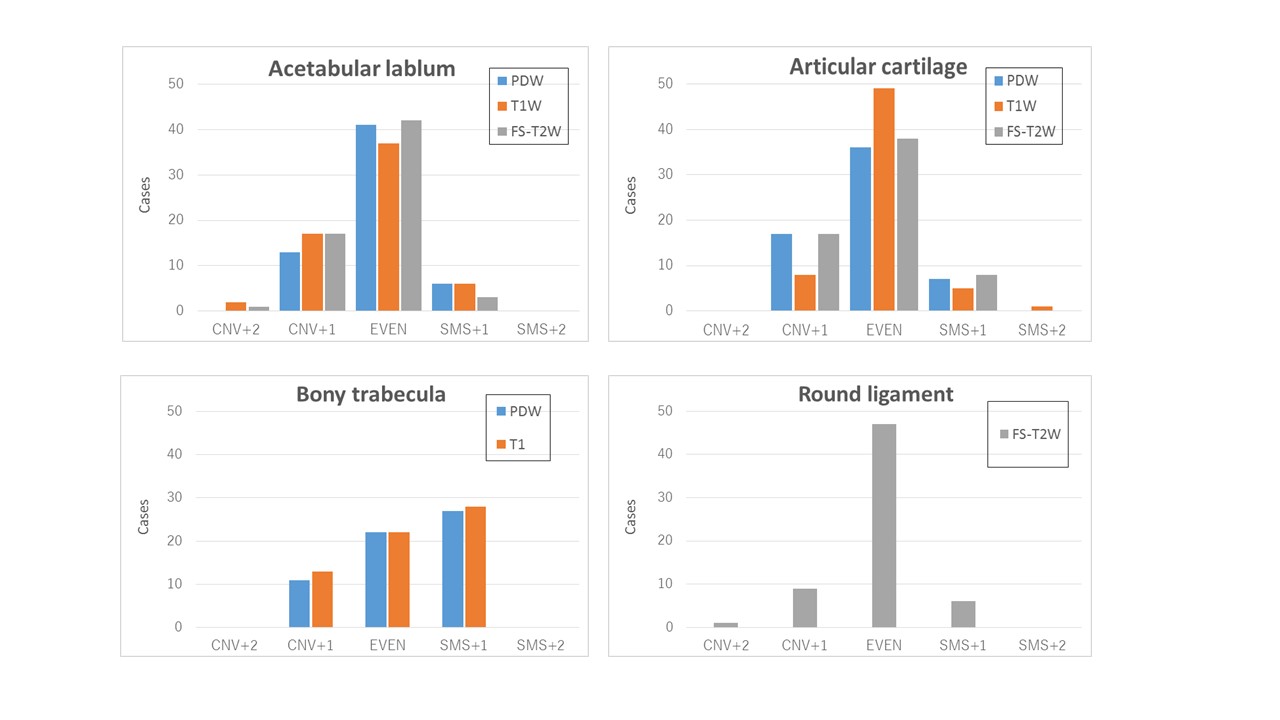
This study was approved by the local ethics committee, and written informed consent was obtained from all patients prior to the MR examinations. MR examination was performed on a 3-T whole-body scanner (MAGNETOM Skyra ; Siemens Healthcare GmbH, Erlangen, Germany), and an 18-channel Body coil. Twenty-one patients (4 males, 17 females, average age of 51.5 years, age range: 22 to 76 years), who had hip pain and were suspected to have acetabular labral damage, underwent unilateral hip joint MR examination. In addition to our routine protocols of oblique coronal proton-density-weighted (PDW) 2D TSE, axial T1-weighted (T1W) TSE and oblique coronal fat-saturated T2-weighted (FS-T2W) TSE, prototype SMS TSE protocols using a prototype SMS TSE were also acquired in the identical geometry (Fig.1 and 2). At the time, system predicted SAR values of each protocol were recorded before the actual scan. therefore there was no actual scan between SAR prediction with and without SMS protocol and they were done in as short a time as possible. A two-sided paired Student’s t-test was used for statistical evaluation. Three board-certificated radiologists evaluated the set of the images with and without SMS for imaging quality of these components: acetabular labrum, articular cartilage, bony trabecula within the femoral neck (only for PDW and T1W), and the round ligament (only for FS-T2W), independently and blindly. The quality was rated using five-point scales; CNV+2 = CNV is superior to SMS, CNV+1 = CNV is slightly superior to SMS, EVEN = CNV is equal to SMS, SMS+1 = SMS is slightly superior to CNV, SMS+2 = SMS is superior to CNV. Inter-observer agreement for the qualitative rating was calculated using Spearman's rank correlation coefficient test. Data analysis was performed with JMP® 12 (SAS Institute Inc., Cary, NC, USA). A p-value of less than 0.05 was considered significant.RESULTS
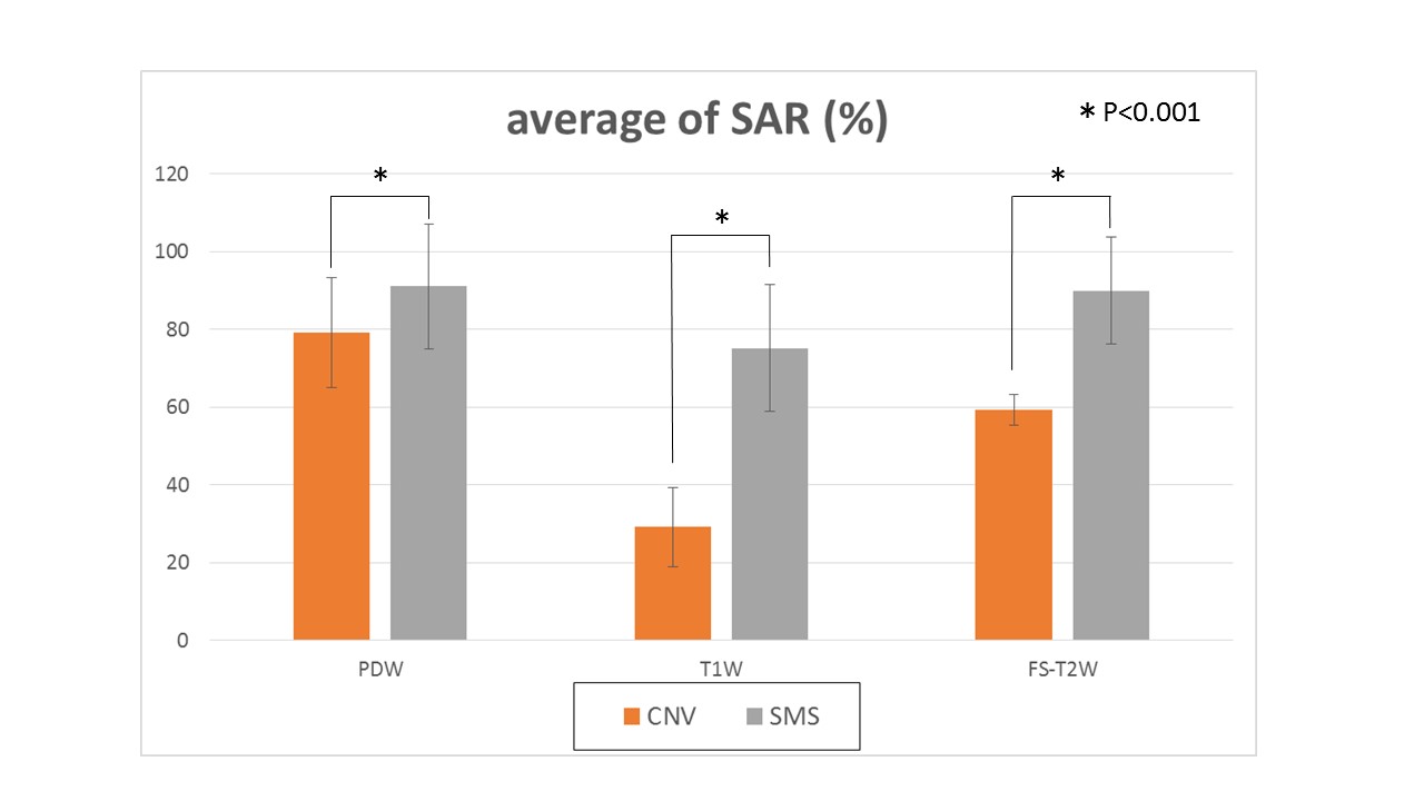
SMS imaging reduced the acquisition time of the total three sequences from 12m16s to 7m35s. Total combined cases of the imaging quality are in Fig. 3. Total 41 cases (68.3%) out of 60 cases were rated as EVEN for delineating the acetabular labrum with PDW, whereas 13 cases (21.7 %) were rated as CNV+1 , 6 cases (10%) as SMS+1, no case was rated as CNV+2 or SMS+2. SMS for delineation of bony trabecular was evaluated slightly better than CNV. No significant correlation in three radiologists scores was found, except for the acetabular labrum with PDW, (rs=0.6023 and p-value = 0.0049). SMS indicated high SAR values of approximately 80-90 % of the simulated limit (Fig. 4). They were significantly higher than CNV (p<0.001 for all sequences).DISCUSSION
SMS indicated relatively equal imaging quality to CNV imaging for delineation of acetabular labrum, articular cartilage, and the round ligament, whereas, slightly better for bony trabecular demonstration. By using SMS, approximately 40% reduction of the acquisition time may provide advantage for clinical MR examinations of the hip joint. Shorter examination time can contribute to increase the examination throughput or be exploited to improve the spatial resolution or SNR.CONCLUSION
With significant reduction of the acquisition time, and with comparable image quality to CNV imaging, SMS can be applied to the clinical MR examination of the hip joint.Acknowledgements
No acknowledgement found.References
1.Kenkel D et al. Whole-Body DiffusionImagingApplying Simultaneous Multi-Slice Excitation. Fortschr Röntgenstr 2016; 188: 381–388
2.David A. Feinberg et al. Ultra-Fast MRI of the Human Brain with Simultaneous Multi-Slice Imaging. J Magn Reson . 2013 April ; 229: 90–100.
3.Crema MD, et al. A narrative overview of the current status of MRI of the hip and its relevance for osteoarthritis research - what we know, what has changed and where are we going? Osteoarthritis and Cartilage (2016),2016 Sep 13. pii: S1063-4584(16)30275-8.
Figures