5022
Application of Simultaneous Multi-Slice TSE in High-Resolution Hand and Foot Imaging1Radiology, Zhengzhou University People’s Hospital, Zhengzhou, People's Republic of China, 2Siemens Healthcare, MR Collaborations NE Asia, Beijing, People's Republic of China, 3Siemens Medical Solutions USA, Inc., MN, United States
Synopsis
The turbo spin echo sequence is one of the most frequently used MR sequences in routine musculoskeletal exams. Its short scan time has allowed it to become a popular clinical choice because it reduces the possibility of motion artifact especially in patients with low tolerance for MR exams. In order to further shorten the scan time and increase temporal and/or spatial resolution, simultaneous multi-slice technique has been widely applied during brain image acquisition. In this study we tried to apply a prototypical SMS-TSE sequence to hand and foot imaging. Compared to the standard TSE sequence, the SMS-TSE had the same image quality and a shorter acquisition time.
Purpose
The turbo spin echo (TSE) sequence is widely used in musculoskeletal imaging due to its ability to provide excellent contrast in cartilage, ligaments, menisci, and periarticular soft tissues. However, its acquisition speed is limited by T2 decay restraints and a loss of signal amplitude when using a large turbo factor. Integrated parallel imaging techniques (iPAT) were developed to accelerate data acquisition using a k-space under-sampling strategy, but the signal-to-noise ratio (SNR) decreases when increasing the iPAT factor to a higher level. The simultaneous multi-slice (SMS) technique is a newly developed acceleration technique using multi-band RF pulses to simultaneously excite, acquire, and reconstruct multiple slices with blipped-CAIPIRINHA,. The purpose of this study was to investigate the feasibility of applying SMS to hand and foot imaging by comparing it with the routine sequences TSE and iPAT in regards to imaging time and images quality.Methods
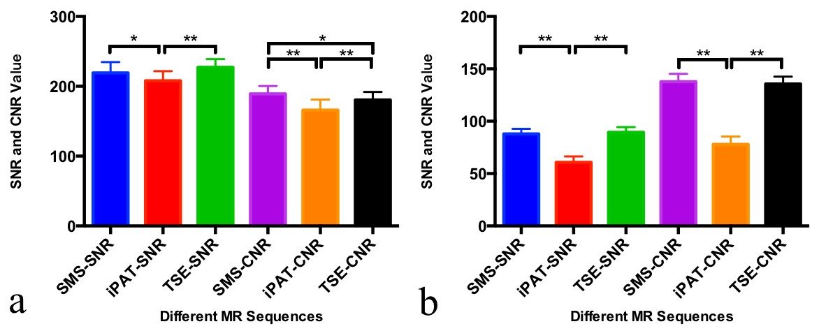
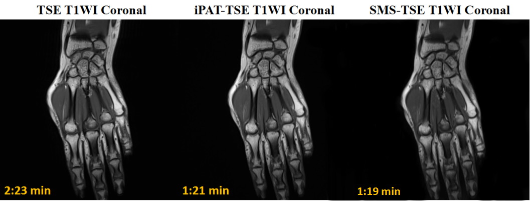
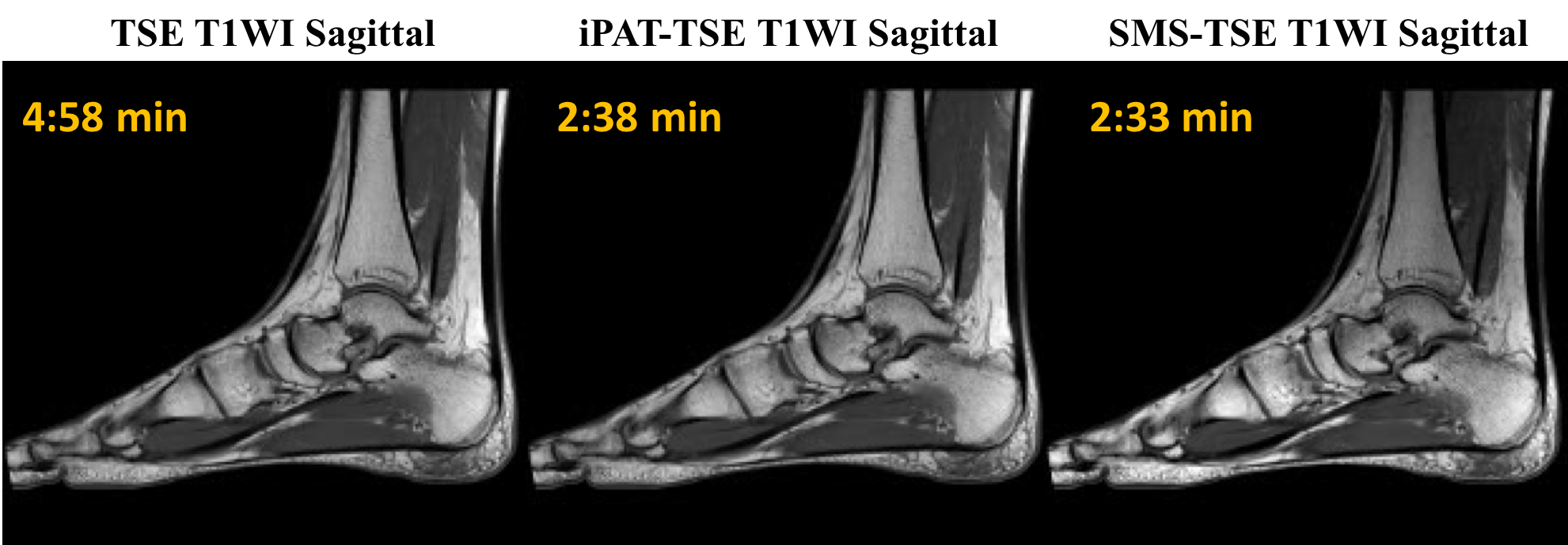
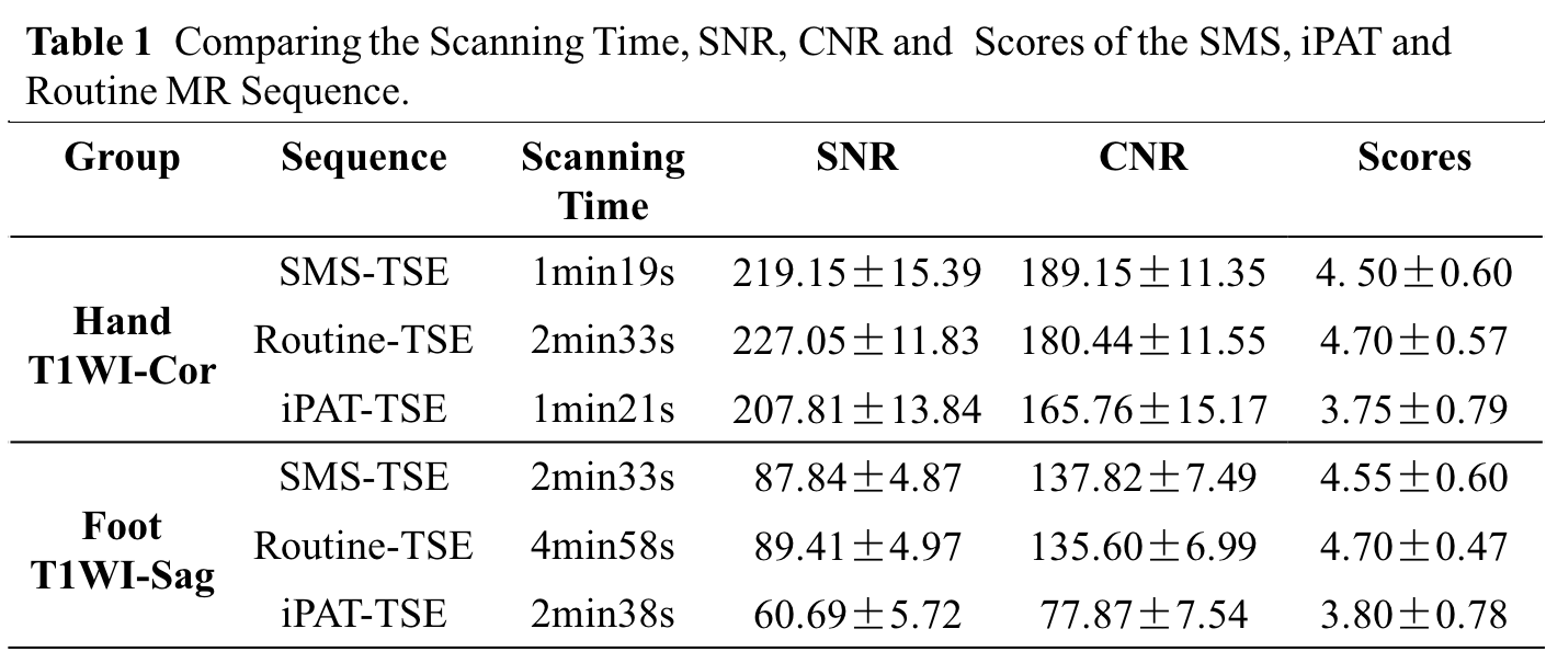
40 patients underwent MR exams on a MAGNETOM Prisma 3T MR scanner (Siemens, Erlangen, Germany). A sixteen-channel foot/ankle coil and a sixteen-channel hand/wrist coil were used. The MR exam included routine TSE, TSE with iPAT (iPAT-TSE), and a prototype SMS-TSE sequence. Images were acquired in axial, sagittal, and coronal planes. The parameters for the hand were: TR/TE=840/11ms for T1WI, 3000/98ms for T2WI, 3000/53ms for PDWI; slice thickness=2.5mm; slices=20; FOV=230×230mm2; matrix=512×358; the parameters for the foot were: TR/TE=600/11ms for T1WI, 3000/38ms for PDWI; slice thickness=3mm; total slice=24; FOV=250×203mm2; matrix=576×328. The iPAT=2 for iPAT-TSE, and the slice acceleration factor=2 for SMS-TSE. The apparent signal-to-noise ratio (SNR) and contrast-to-noise ratio (CNR) were calculated for the MR images collected by the T1WI-Cor of the hand and the T1-sag of the foot. The background areas outside the foot and hand were selected as one big ROI to estimate the noise. Although parallel imaging technique such iPAT and SMS induce spatially varying g-factor and noise, the large spatial support of the ROI used for noise estimation should mitigate this effect. Image quality was rated by radiologist blindly based on a five-point evaluation scale as follows: 5 = uniform signal, excellent contrast, no artifacts; 4 = mild signal inhomogeneity, good contrast, mild artifacts; 3 = moderate signal inhomogeneity, moderate contrast, moderate artifacts; 2 = strong signal inhomogeneity, poor contrast, prominent artifacts; 1 = non-diagnostic.The SNR and CNR were statistically analyzed using LSD-t test. Image quality scores were tested for statistical significance by using the Games-Howell test.Results
Compared with the Routine-TSE and iPAT-TSE sequences, the SMS-TSE sequence could greatly shorten the scan time (Table 1) to 55% of the standard TSE. For hand and foot MR imaging, the SNR,CNR of the SMS-TSE sequence are close to the Routine-TSE and slightly higher than the iPAT sequence. No significant difference was observed between the SMS-TSE and Routine-TSE sequence for the SNR(Figure 1). For the hand MR imaging, the image quality scores of the SMS-TSE and Routine-TSE sequences were significantly higher than that of the iPAT sequence(Hand: p=0.005, SMS vs iPAT; p=0.001, Routine vs iPAT; p=0.536, Routine vs SMS). The results of image quality scores of the foot MR image is similar to the hand MR imaging(Foot: p=0.004, SMS vs iPAT; p=0.001, Routine vs iPAT; p=0.659, Routine vs SMS) . With reduced total acquisition time, the SMS-TSE sequence demonstrated similar image quality as a routine TSE sequence in T1WI (Figure 2 and 3) .Discussion
We found that both iPAT and SMS can reduce scan time significantly; however, the SNR and CNR of the images obtained with iPAT were lower than those obtained with SMS due to the under-sampling of the k-space. The SNR of SMS-TSE and of Routine-TSE were comparable, thus indicating that SMS-TSE could significantly reduce the scan time for foot and hand imaging when acquiring thin slices with large coverage. Since SMS-TSE mainly reduces the TR for image acquisition, its full potential may not be realized in cases where small coverage or a limited number of slices is used.Conclusion
The SMS-TSE sequence can be used to obtain high quality images for the hand and foot at 3T with 45% less acquisition time than the standard TSE sequence.Acknowledgements
No acknowledgement found.References
[1] Wang Y, et al. Neuroimage. 2015 ,113:279-88.
[2] Setsompop K, et al. MRM. 2012May;67(5):1210-24.
[3] Moeller S, et al. MRM. 2010 May;63(5):1144-53.
[4] Setsompop K, et al. Neuroimage. 2012 Oct 15;63(1):569-80.
[5] Breuer FA,et al. MRM. 2005 Mar;53(3):684-91.
Figures