5020
Compressed Sensing 3D GRASE for Faster PD-weighted Knee Imaging1Department of Radiology and Nuclear Medicine, Erasmus MC, Rotterdam, Netherlands, 2GE Healthcare B.V., Hoevelaken, Netherlands
Synopsis
Accelerated 3D-FSE using Compressed Sensing (CS) with a 30% scanning time reduction has been recently introduced and evaluated for knee MR images. 3D-GRASE is a hybrid FSE-EPI sequence that can achieve higher time-efficiency scans, since it acquires more k-space data per refocusing pulse. The purpose of this work is to present CS 3D-GRASE to achieve even faster musculoskeletal MRI acquisitions.
Introduction
Three-dimensional musculoskeletal MRI has been proved to provide high-spatial resolution and high-quality multi-planar reformats, enabling detection of more subtle injuries1. Nevertheless, their clinical use is restricted due to their long acquisition times2. Recently, accelerated 3D-FSE using Compressed Sensing (CS) with a 30% scanning time reduction has been introduced and evaluated for knee MR images2. 3D-GRASE3 is a hybrid FSE-EPI sequence that can achieve higher time-efficiency scans, since it acquires more k-space data per refocusing pulse. The purpose of this work is to present CS 3D-GRASE to reduce scan time in musculoskeletal MRI.Methods
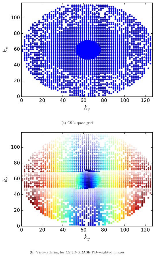
CS k-space grid is generated in a sequential order, combining Parallel Imaging (PI) + CS, as previously described for CS-FSE4. A fully-sampled elliptical grid is regular undersampled according to the pattern needed by ARC5. Then, a Gaussian pseudo-random subsampling is applied except for the autocalibration area needed for the Autocalibrating Reconstruction for Cartesian sampling (ARC) algorithm. Figure 1a shows the final CS k-space grid for 3D-GRASE and 3D-FSE. Two view-ordering techniques were combined to enable CS+PI in 3D-GRASE: SORT6, which splits off-resonance and T2 effects in different phase encoding directions, and linear modulation7, which mapping the signal modulation into k-space to include 2D acceleration, achieve different image contrasts and mitigate artifacts.
The algorithm uses any k-space sampling pattern, as list of k-space positions, the Echo train length (ETL) and EPI factor (N) as inputs. To each k-space position it assigns a train number, echo number along the ETL, and the echo in N. To obtain PD-weighted contrast, the center of the k-space has to be filled at the beginning of the Echo Train (ET). This is accomplished by separating the positions with $$$k_y \geq k_{y\_center}$$$ and $$$k_y<k_{y\_center}$$$ and sorting on $$$k_z$$$ each sublist. Subsequently, each sublist is split in N equally sized parts, where list number defines then echo in N. Each sublist is sorted on k_y and split in ETL (specifying echo number), with each sorted on $$$k_z$$$. The position in the final list specifies the train number, except that the $$$k_y \geq k_{y\_center}$$$ and $$$k_y<k_{y\_center}$$$ lists are interleaved along the acquisition (See Figure 1b).
Additionally one reference echo train without playing out slice and phase gradients is acquired for phase and amplitude correction8. Echoes were time corrected by shifting the echo peak to the center of the time acquisition window. Phase correction was performed by point-by-point subtraction of the phases of the Fourier transformed reference echoes from the Fourier transformed echoes in the acquisition. Subsequently, skipped random samples were reconstructed by minimizing the total variation for each channel using a conjugate gradient minimizer obtaining a k-space with the regular undersampling needed for ARC. Then, ARC was applied to obtain the final fully k-space per channel. Finally, they were combined by sum of squares and correction for gradient non-linearities was applied to each slice.
Results
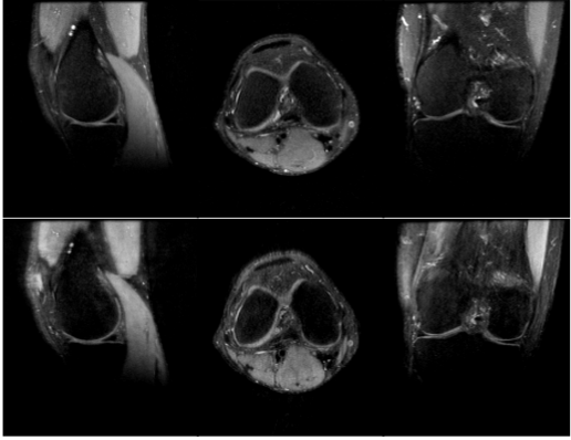
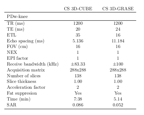
Human in vivo experiments were performed on a 3T GE MR750 scanner with a four channel transmit-receive knee coil for whole knee PD-weighted images. Table 1 shows the acquisition parameters and time and values for CS 3D-GRASE and CS 3D-FSE in a knee. The parameters used for GRASE acquisition were chosen to maintain as much as possible the same TEeff as FSE. Figure 2 shows three orthogonal slices of PD-weighted knee acquired with CS 3D-FSE and the proposed CS 3D-GRASE.
Discussion/Conclusions
The presented view-ordering scheme for 3D-GRASE has demonstrated to be compatible with CS acquisitions. For the same acceleration factor, a high reduction in time (~25%) is obtained with CS 3D-GRASE compared to CS 3D-FSE for PD-weighted knee images. The three orthogonal slices visually show a slight blur with no artifacts or distortions. A modest difference in contrast can be appraised between CS 3D-GRASE and CS 3D-FSE, due to the slight difference in the TEeff value. Further clinical evaluation is needed in order to assess the clinical application of CS 3D-GRASE.Acknowledgements
This work was supported by a GE grant. We would like to thank Rob Peters, Hendrik de Leeuw, Gyula Kotek and Piotr Wielopolski.References
[1] Naraghi, A., & White, L. M. (2012). Three-dimensional MRI of the musculoskeletal system. American Journal of Roentgenology, 199(3), W283-W293.
[2] Kijowski, R., Rosas, H., Samsonov, A., King, K., Peters, R. and Liu, F. (2016), Knee imaging: Rapid three-dimensional fast spin-echo using compressed sensing. J. Magn. Reson. Imaging.
[3] Koichi Oshio and David A Feinberg. GRASE (gradient-and spin-echo) imaging: A novel fast MRI technique. Magnetic Resonance in Medicine, 20(2):344–349, 1991.
[4] Kijowski, R., Rosas, H., Samsonov, A., King, K., Peters, R. and Liu, F. (2016), Knee imaging: Rapid three-dimensional fast spin-echo using compressed sensing. J. Magn. Reson. Imaging.
[5] PJ Beatty, AC Brau, S Chang, SM Joshi, CR Michelich, E Bayram, TE Nelson, RJ Her- fkens, and JH Brittain. A method for autocalibrating 2-D accelerated volumetric parallel imaging with clinically practical reconstruction times. In Proc Intl Soc Magn Res Med, volume 15, page 1749, 2007.
[6] John P. Mugler. Improved three-dimensional GRASE imaging with the sort phase-encoding strategy. Journal of Magnetic Resonance Imaging, 9(4):604–612, 1999.
[7] Reed F. Busse, Anja C.S. Brau, Anthony Vu, Charles R. Michelich, Ersin Bayram, Richard Kijowski, Scott B. Reeder, and Howard A. Rowley. Effects of refocusing flip angle modulation and view ordering in 3D Fast Spin Echo. Magnetic Resonance in Medicine, 60(3):640– 649, 2008.
[8] Jorge Jovicich and David G. Norris. GRASE imaging at 3 Tesla with template interactive phase–encoding. Magnetic Resonance in Medicine, 39(6):970–979, 1998.
Figures