5018
Compressed Sensing SEMAC MRI of Total Knee Arthroplasty Implants: Intra-subject Comparison at 1.5 and 3 Tesla1The Johns Hopkins University School of Medicine, Baltimore, MD, United States, 2Orthopaedic University Hospital Balgrist, 3Siemens Healthcare USA, 4Siemens Healthcare GmbH
Synopsis
Because susceptibility artifacts increase with field strength, MRI of knee arthroplasty implants is commonly performed at 1.5T. However, 3T MRI offers substantially higher SNR and may be the only available option. SEMAC can achieve substantial metal artifact reduction at 3T, but requires prolongs scan times. Capitalizing on the inherent sparsity of SEMAC data, compressed sensing-based pseudo-randomized undersampling and iterative reconstruction can substantially accelerate data acquisition. We show the clinical feasibility of highly accelerated 3T MRI of CoCr knee arthroplasty implants using a compressed sensing SEMAC TSE sequence with metal artifact reduction capabilities and acquisition times similar to 1.5T.
Introduction
Because susceptibility artifacts increase with field strength, MRI of arthroplasty implants is most commonly performed at 1.5T. However, 3T MRI has become popular due to the increased signal-to-noise ratio and may be the only available option at some sites. Slice-encoding metal artifact correction (SEMAC) has the potential to substantially reduce metal artifacts at 3T [1]. The required higher number of SEMAC encoding steps at 3T, however, results in long acquisition times, which is exacerbated by specific absorption rate limitations. The inherent sparsity of SEMAC can be exploited for compressed sensing-based acceleration of data acquisition through pseudo-randomized undersampling and iterative reconstruction [2].Purpose
We prospectively compared highly accelerated compressed sensing SEMAC MRI of knee arthroplasty implants at 1.5T and 3T within the same subjects.Methods
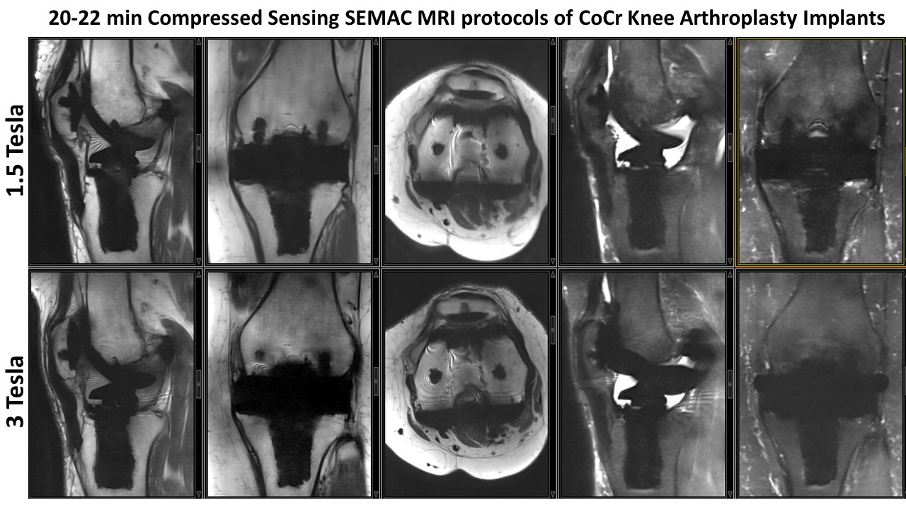
Ten subjects with Cobalt-Chromium-containing knee arthroplasty implants prospectively underwent MRI at 1.5T (MAGNETOM Aera, Siemens Healthcare, Erlangen, Germany) and 3T MRI (MAGNETOM Skyra, Siemens Healthcare) using 15-channel transmit-receive knee coils. Compressed sensing SEMAC acceleration was realized with a prototype implementation of 8-fold incoherent undersampling of the 2D phase-encoding matrix and non-linear, SENSE-type reconstruction with L1-norm-based regularization [3]. At 1.5T, sagittal, coronal and axial intermediate-weighted (IW) as well as sagittal and coronal STIR compressed sensing SEMAC images were acquired (IW: TR 3750 ms, TE 28 ms, pixel size 0.5 x 0.5 mm2, SL 3-4 mm; STIR: TR 4500 ms, TE 7.5 ms, TI 160 ms, pixel size 0.6 x 0.6 mm2, SL 4 mm) with 15 SEMAC-encoding steps and total acquisition time of 19:49 min. At 3T, the same sequences were acquired (IW: 3800 ms, 33 ms, 0.4 x 0.4 mm2, 3 mm; STIR: 5400 ms, 8.2 ms, TI 220 ms, 0.5 x 0.5 mm2, 4 mm) and 21 SEMAC-encoding steps and a total acquisition time of 21:42 min. Two fellowship-trained musculoskeletal radiologists graded metal artifact reduction, edge sharpness, noise, fat suppression, and visibility of bone, arthroplasty-bone interface, and soft tissue structures using standardized Likert scales. Non-parametric tests were used. P< 0.05 was considered significant.Results
All subjects successfully underwent both MRI examinations at 1.5T and 3T (Figure 1). Compared to compressed sensing SEMAC at 1.5T, compressed sensing SEMAC at 3T was characterized by a mildly larger metal artifacts (p<0.05), sharper images (p<0.05), less noise (p<0.05), similar fat suppression (p=0.592), similar visibility of bone (p=0.123), mildly inferior implant-bone interface visibility (p=0.045) and superior display of soft tissues (p<0.05).Discussion
We demonstrate the clinical feasibility of compressed sensing SEMAC for MRI around cobalt-chromium knee arthroplasty implants at 1.5 and 3 Tesla field strength. The intrinsically higher signal-to-noise ratio of 3T can be utilized to achieve higher spatial resolution. Despite the use of a higher number of SEMAC-encoding steps to address the increased magnitude of the metal artifact at 3T, scan times for each CS-SEMAC pulse sequence could be kept well below 5 minutes. Thus, a substantial reduction of the metal artifact was achieved, which was only marginally larger than metal artifacts in the same subjects at 1.5T.Conclusion
CS-SEMAC MRI facilitates MRI around knee arthroplasty implants at 1.5 and 3T, with mildly larger metal artifacts at 3T. Complete MRI exam of a subjects with cobalt-chromium knee implants can be performed in 25 min or less with both field strengths.Acknowledgements
The authors would like to acknowledge Jens Wetzl and Michael Zenge for their work on the image reconstruction framework.References
1. Lu W, Pauly KB, Gold GE, Pauly JM, Hargreaves BA. SEMAC: Slice Encoding for Metal Artifact Correction in MRI. Magn Reson Med. 2009 Jul; 62(1): 66–76.
2. Fritz J, Ahlawat S, Demehri S, Thawait GK, Raithel E, Gilson WD, Nittka M. Compressed Sensing SEMAC: 8-fold Accelerated High Resolution Metal Artifact Reduction MRI of Cobalt-Chromium Knee Arthroplasty Implants. Invest Radiol. 2016 Oct;51(10):666-76.
3. Liu j, Rapin J, Chang T, Schmit P, Bi X, Lefebvre A, Zenge M, Mueller E, Nadar M. Regularized reconstruction using redundant Haar wavelets: A means to achieve high under-sampling factors in non-contrast-enhanced 4D MRA. ", Proceeding of the International Society for Magnetic Resonance in Medicine, 20TH Annual Meeting and Exhibition, Melbourne, Australia, 5-11 May 2012, Vol. 20, 21 April 2012 (2012-04-21), pages 2237.
Figures