5016
Knee-to-coil automatic distance detection for misalignment alert system during MRI acquisitionTakamasa Sugiura1, Toshimitsu Kaneko1, Tomoyuki Takeguchi1, Kensuke Shinoda2, Takuya Fujimaki2, and Hiroshi Takai2
1Toshiba Corporation, Kawasaki, Japan, 2Toshiba Medical Systems Corporation, Otawara, Japan
Synopsis
For high quality knee MRI image acquisition, the coil must be centrally aligned with the knee. However, precise alignment can suffer from patient motion and is currently performed by eye. We propose a method to automatically measure the misalignment between coil and knee to alert the clinical operator. This is done by calculating the distance between the coil and the knee joint gap by processing the localizer image with a machine learning technique, which was achieved with a mean accuracy of 3.3 mm. Our experiments further indicated a safe margin for knee-to-coil misalignment within a threshold of 20 mm.
Purpose
A knee MR scan is indispensable for the diagnosis of tears of ligaments and meniscus. To take a high quality image, the knee coil must be aligned correctly. In particular, if the knee center is far from the center of coil, the resultant knee image will suffer from low contrast partially due to the low coil sensitivity range. Figure 1 shows the effects on image quality as the knee/coil distance is increased. As coils are currently aligned manually by the operator, errors can arise both from such manual alignment as well as from patient movements. Therefore, we propose an alert system to instruct the operator to adjust the coil position. The system takes a fast localizer image at a low resolution, from which the distance between the knee joint gap and coil center is automatically calculated. If the distance is large, precise instructions are displayed, e.g. “Shift 4 cm towards head”, so that the coil is appropriately aligned. In the setup, we assume the coil center to be aligned with the magnetic field, which can be easily carried out with the aid of the guiding beam. To the best of the authors’ knowledge, this is the first alert system for knee MR alignment purposes.Methods
Figure 2 shows the illustration of the proposed method. In the workflow, a 3D fast field echo (FFE) anisotropic localizer image covering both the left and right knee is acquired without slice positioning using a 3T MRI scanner with FOV = 500 × 500 × 160 mm in less than 25 seconds. Note that the center of image is at the center of coil because of the absence of slice positioning. The knee center is detected using extremely randomized trees1, which is a fast and highly robust classification technique. Then, the distance between coil and knee is calculated as the distance from the center of image to the detected center of knee in the head-foot direction.Results and Discussion
Our experiments used 30 localizer images from patients. Ground truths of knee centers were manually identified by two experienced operators. Mean and standard deviation of the distance error with the proposed method was 3.3 ± 2.5 mm, while the inter-technologists error was 3.8 ± 3.5 mm, so the knee center was concluded to have been detected accurately. We also obtained the operator’s subjective assessment of whether coil position adjustment is necessary based on the observation of the localizer images. Figure 3 shows the results of detected distance together with operator assessment, which shows a clear direct correlation between the necessity for position adjustment and the detected distance. Our results suggest that an alert function is appropriate to instruct operators when distances over 20 mm are estimated, based on our qualitative assessment from skilled operators.Conclusion
We proposed an automatic knee-to-coil distance measurement system to alert MR operators of misalignments in the coil setup, based on an automatic knee joint gap detection from localizer images using machine learning techniques. Experimental results showed that our measurements are accurate and are highly correlated with the operators’ own quality assessment. By alerting the operator of misalignments in excess of 20 mm, it is possible to communicate the necessary adjustments and obtain better quality MR images.Acknowledgements
We would like to express our gratitude to the technologists of Kameda Medical Center for clinical advice and data correction. We also thank Dr. Marco Visentini-Scarzanella for proofreading.References
1. Pierre G, Damien E, Louis W. Extremely randomized trees. Machine Learning. 2006;63(1):3-42.Figures
Figure
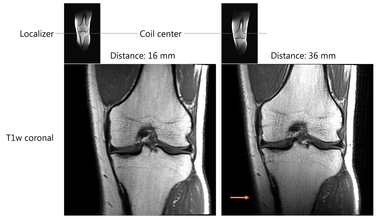
1: Examples of decreasing of image quality due to the knee-to-coil distance.
Localizer and T1w coronal images in each case. Left: distance is 16 mm,
resulting in a good quality T1w coronal image. Right: with a distance of 36 mm
the image contrast decreases towards the bottom of the image (orange arrow).
Figure
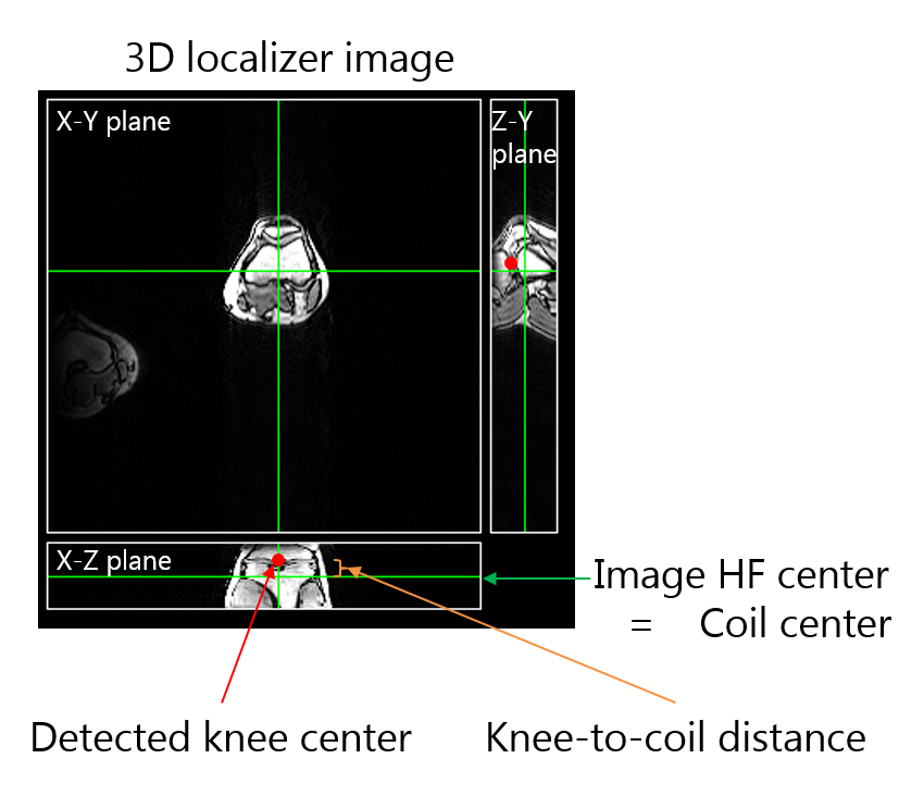
2: Illustration of our method. Green crossing lines on the 3D anisotropic
localizer image show the position of the cross-section planes (X-Y, X-Z, Z-Y). The
horizontal green line on the X-Z plane (equivalent to the vertical line on the
Z-Y plane) is at the coil center. The knee-to-coil distance is calculated as
the distance of the detected knee center (red point) and the coil center.
Figure
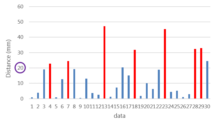
3: Detected distance for each data sample. Red bars are samples where the coil
has to be adjusted according to the skilled operators’ opinion. From the graph,
a distance threshold of 20 mm for the alert system is adequate.