5012
Magnetic resonance imaging estimates of intermuscular fat density in the thigh in sarcopenia population: correlation with physical performances1Institute of Geriatrics and Active Ageing, Tan Tock Seng Hospital, Singapore, Singapore, 2Geriatric Medicine, Tan Tock Seng Hospital, Singapore, Singapore, 3Diagnostic Radiology, Tan Tock Seng Hospital, Singapore, Singapore
Synopsis
Emerging evidence suggests that intermuscular fat (IMF) accumulation is associated with reduced muscle quality and increased risk of physical limitation. However, the impact and mechanism of IMF in sarcopenia or sarcopenic obesity (SO) are still unclear. MRI is a promising tool for early detection of sarcopenia and SO. This may translate to use in clinical trials and in future clinical practice, where quantitative assessment may become standard of care. . This study aims to study a new index that we term “IMF density”, shows promise as an important quantitative variable that reflects patients’ physical performances.
Introduction
Muscle and fat volume is particularly relevant to management of sarcopenia and sarcopenic obese (SO) patients. Magnetic resonance imaging (MRI) is a promising imaging modality to quantify muscle and fat volume including inter-muscular fat (IMF), given its ionizing radiation-free nature and excellent soft tissue contrast. Emerging evidence suggests that IMF accumulation is associated with reduced muscle quality and increased risk of physical limitation. However, the impact and mechanism of IMF in sarcopenia or sarcopenic obesity (SO) are still unclear. This study aims to study a new index which we term “IMF density”, defined as the ratio between IMF and the combination of IMF and muscle volume. We assessed the correlation between the different thigh compartments, including IMF density and subjects’ physical performances.Methods and Materials
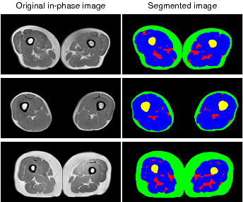
MRI scans of the thigh of 190 healthy community dwelling older adults (age 50-99, average age: 67.85±7.90, 58 males and 132 females) were acquired and analyzed as a part of a larger longitudinal study. Subjects were categorized into four subgroups based on Asian established criteria1: normal, obese, sarcopenia, and SO. Dixon GRE sequences were acquired and muscles, subcutaneous fat (SF), and inter-muscular fat (IMF) were automatically segmented in both thighs using an in-house machine learning based segmentation method2. Volumes of muscles and fat were calculated for the middle third of each patients’ thigh. The IMF density was computed as: VIMF/(VIMF+VMuscle). All the volumes were adjusted by subjects’ height square. Knee extension strength, grip strength, gait speed based on the time to walk 3 m, chair stand, and the Short Physical Performance Battery (SPPB)3 was used to measure physical performances. Spearman correlation coefficients (CCs) between thigh component volumes and physical performances were analysed using SPSS.Results and discussion
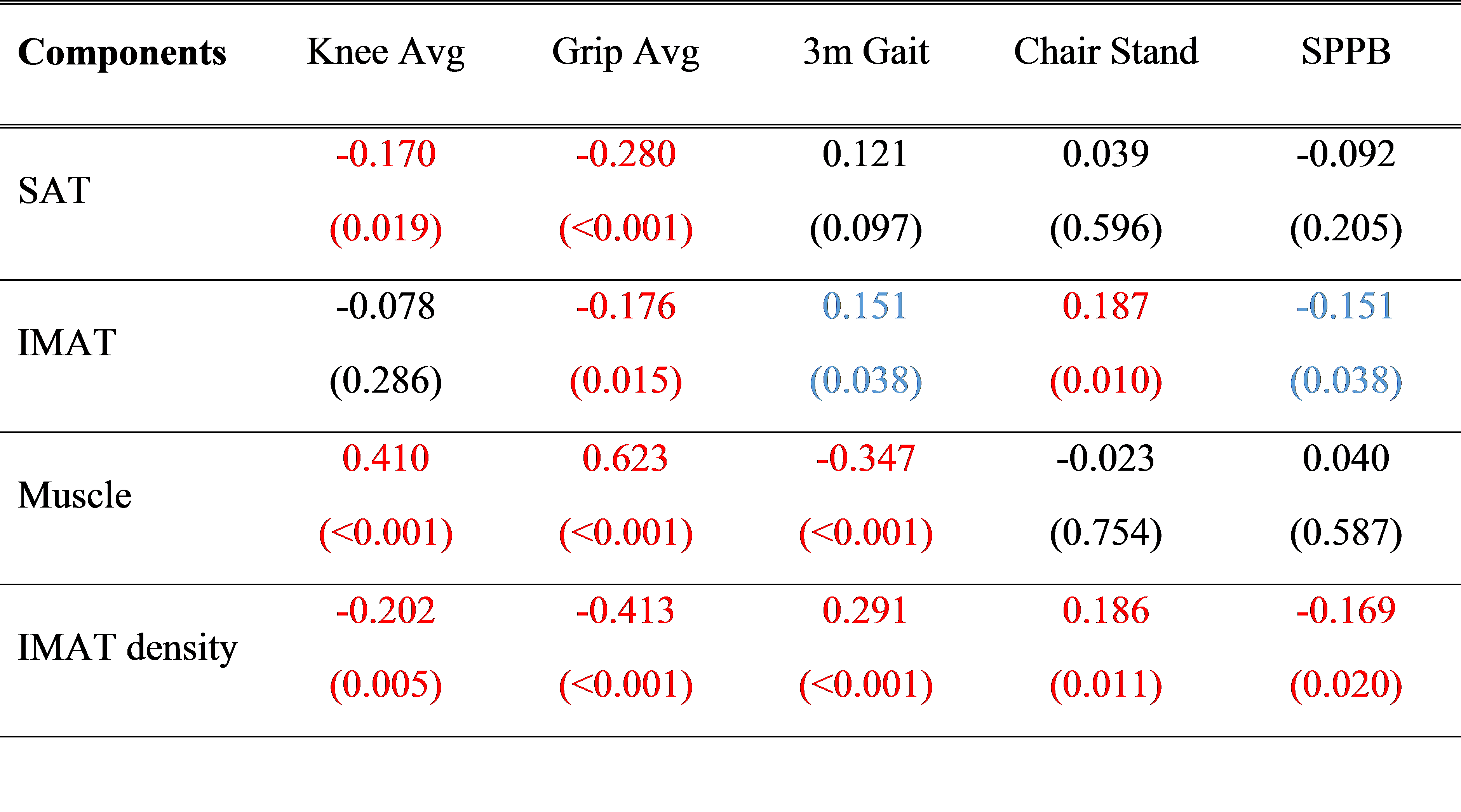
Figure 1 shows sample segmented images of muscle, subcutaneous fat (SF), and IMF in thigh. Spearman correlation coefficients (CCs) between thigh component volumes and physical performances are listed in Table 1 with p values in parentheses. Significant results with p < 0.025 are marked in red and with p<0.05 are marked in blue. The newly introduced IMF density was the only thigh component that correlated significantly with all physical performances (p<0.025); while the muscle and IMF volume alone only correlated with some but not all of the physical performance measurements. The IMF density could be the component that interpret the muscle quality in thigh. In future work, changes of thigh components will be monitored in the longitudinal study. The results showed that detailed analysis of body components using MRI is necessary.Conclusion
MRI is a promising tool for early detection of sarcopenia and SO. This may translate to use in clinical trials and in future clinical practice, where quantitative assessment may become standard of care. IMF density shows promise as an important quantitative variable that reflects patients’ physical performances.Acknowledgements
No acknowledgement found.References
1. Chen, Liang-Kung, Li-Kuo Liu, Jean Woo, Prasert Assantachai, Tung-Wai Auyeung, Kamaruzzaman Shahrul Bahyah, Ming-Yueh Chou et al. "Sarcopenia in Asia: consensus report of the Asian Working Group for Sarcopenia." Journal of the American Medical Directors Association 15, no. 2 (2014): 95-101.
2. Yang YX, Chong MS, Tay L, Yew S, Yeo A, Tan CH (2016) Automated assessment of thigh composition using machine learning for Dixon magnetic resonance images. Magn Reson Mater Phys, Biol Med:1-9. doi:10.1007/s10334-016-0547-2
3. Guralnik, Jack M., Eleanor M. Simonsick, Luigi Ferrucci, Robert J. Glynn, Lisa F. Berkman, Dan G. Blazer, Paul A. Scherr, and Robert B. Wallace. "A short physical performance battery assessing lower extremity function: association with self-reported disability and prediction of mortality and nursing home admission." Journal of gerontology 49, no. 2 (1994): M85-M94.
Figures