5001
Development of an MR-Compatible Ergometer for Use in Quantifying Human Skeletal Muscle Bioenergetics During Supine Dynamic Contractions of the Knee Extensors1Human Magnetic Resonance Center, Institute for Applied Life Sciences (IALS), University of Massachusetts, Amherst, MA, United States, 2Department of Mechanical & Industrial Engineering, University of Massachusetts, Amherst, MA, United States, 3Kinesiology, University of Massachusetts, Amherst, MA, United States
Synopsis
The goal of this project was to develop an MR-compatible, multi-modal ergometer for the reliable measurement of human skeletal muscle torque, velocity, power and joint angle during 31P MRS studies of knee extensor muscle energetics. Intracellular [PCr], [Pi] and pH were determined in the vastus lateralis with 4-s time resolution during 4 min of maximal voluntary isokinetic contractions at 240 degrees per second, with a 30 degree range of motion. High S/N for both the MRS and power data indicate that this tool will be useful in future studies of in vivo muscle bioenergetics.
Introduction
For more than 30 years, phosphorus MR spectroscopy (31P MRS) has been a reliable and robust tool for noninvasive investigation of human muscle bioenergetics 1. 31P MRS can be used to quantify the concentration and kinetics of high-energy phosphorus metabolites and pH in resting muscle, as well as during and following muscular work. For example, the rate constant for phosphocreatine (PCr) recovery can be used to quantify the muscle’s capacity for oxidative energy (ATP) production by the mitochondria 2, 3. One advantage of this method is that measures of muscle torque or work are not necessary. In contrast, accurate determination of the ATP cost of contraction (work per ATP), including ATP production by the creatine kinase reaction, nonoxidative glycolysis and oxidative phosphorylation 4-6, requires precise measures of muscular work. However, commercial availability of MR-compatible ergometers is very limited, as are the capabilities of these devices. Therefore, the goal of this project was to develop and test a new ergometer for measurement of human muscle torque, velocity, joint angle and power during knee and full-leg extension exercise during simultaneous measures of 31P MRS in the 3T environment.Materials and Methods
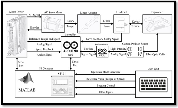
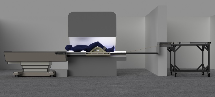
The ergometer was designed to quantify torque and power during isometric, isotonic and isokinetic knee and full-leg extension exercise, with the muscle of interest positioned in the isocenter of a 70-cm bore Skyra 3T system (Siemens Medical Systems, Germany). Results from pilot studies of a knee extension protocol are presented here. Required output variables included position (to determine knee angle), torque and velocity data with high S/N that would not contribute noise to 31P MRS signals obtained at rapid (~2s) repetition rates. The test protocol consisted of 4 min of maximal voluntary isotonic contractions at 240 dps, repeated once every 2 s, which was designed to induce fatigue 7. Phosphorus spectra were acquired before, during and for 10 min following the protocol. Prior to the exercise study, a 500-ml phosphoric acid phantom was used to determine the temporal stability of the 6 x 8 cm 31P elliptical loop transmit/receive RF surface coil (J. Smith, University of Oregon, Eugene OR, USA). Subsequently, this coil was positioned over the vastus lateralis muscle and held in place with an elastic wrap and velcro straps. Gradient-echo scout images and T1-weighted axial images along the femur were used to confirm optimal positioning of the leg in the isocenter, and correct positioning of the coil over the muscle. Magnetic field homogeneity was optimized on water using the body coil, and confirmed on the PCr peak of the 31P signal to yield width at half-height of ~14 Hz. 31P-MRS data were collected in 3 healthy volunteers (mean age 24.5 years). Spectra were acquired using a pulse-and-acquire free induction decay sequence using adiabatic half passage pulses. The parameters included: 4000 Hz band width, 2048 complex points, 2s repetition time. Spectra were averaged as follows: 60s at rest, 4s during contraction protocol, and 4-30s during recovery. Spectra were processed using jMRUI version 5.2 and quantified using a non-linear least squares algorithm 8.Resulst and Discussion
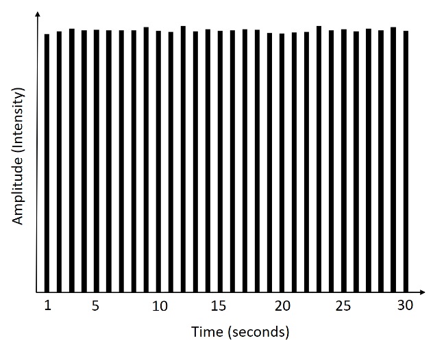
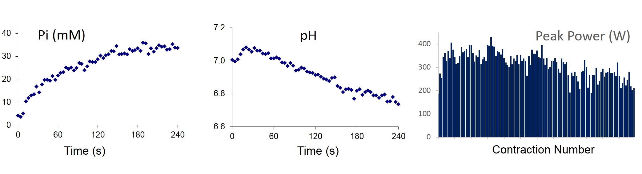
Design and testing of the ergometer resulted in a system well-suited to the 70-cm bore of the 3T (Figures 1 and 2). The system was made MR-compatible by using non-magnetic materials in the bore and locating all of the powered elements away from the field (Figure 2). Powered actuation elements were connected mechanically through a system of low-stretch cables and pulleys to passive components inside the scanner. The ergometer applies controlled resistive loads on the lower limb, enabling study of the muscle while it is near its physical workload limits. A 30 degree range of motion (ROM), from 105 to 135 degrees, was obtained. The closed-loop force control allowed for isometric, isotonic and isokinetic contractions; fatigue was produced using the isokinetic mode. The temporal stability of the 31P coil was good, as evidenced by a low coefficient of variation (0.7%) for signal intensity over the 30-s data collection period (Figure 3). The contraction protocol produced substantial changes in [Pi] and pH, two known inhibitors of contractile function in muscle 9, and the accompanying decrement in peak power is shown in Figure 4.Conclusion
The custom engineering and construction approach applied here enables collection of high S/N 31P metabolites and pH during maximal work of the human knee extensor muscles. The capacity of this design to control load and velocity during isotonic and isokinetic contractions will advance the study of in vivo muscle energetics by enabling a wide range of contraction protocols.Acknowledgements
No acknowledgement found.References
1. Chance B, Leigh JS, Clark BJ, et al. Control of oxidative metabolism and oxygen delivery in human skeletal muscle: a steady-state analysis of the work/energy cost transfer function. Proc. Natl. Acad. Sci. 1985; 82(24). 8384-8388
2. Meyer RA. A linear model of muscle respiration explains monoexponential phosphocreatine changes. Am. J. Physiol. Cell. Physiol. 1988; 254(4). C548-C553
3. Lanza IR, Bhagra S, Nair KS, et al. Measurement of human skeletal muscle oxidative capacity by 31P-MR spectroscopy: A cross-validation with in vitro measurements. J. Magn. Reson. Imaging. 2011; 34(5) 1143-1150
4. Boska M. Estimating the ATP cost of force production in the human gastrocnemius/soleus muscle group using 31P MRS and 1H MRI. NMR Biomed. 1991; 4(4):173-81
5. Christie AD, Tonson A, Larsen RG, et al. Human skeletal muscle metabolic economy in vivo: effects of contraction intensity, age, and mobility impairment. Am J Physiol Regul Integr Comp Physiol. 2014; 307(9):R1124-35
6. Layec G, Hart CR, Trinity JD, et al. Skeletal muscle work efficiency with age: the role of non-contractile processes. Clinical Science 2015; 128, 213–223.
7. Callahan DM & Kent-Braun JA. Effect of old age on human skeletal muscle force-velocity and fatigue properties. J Appl Physiol. 2011;111, 1345-1352
8. Stefan D, Di Cesare F, Andrasescu A, et al. Quantitation of magnetic resonance spectroscopy signals: the jMRUI software package. Meas Sci Technol 2009; 20(10):104035(9pp).
9. Kent-Braun JA, Fitts RH, Christie AD. Skeletal muscle fatigue. Compr Physiol. 2012; 2:997-1044
Figures