5000
MRI/S Assessment of Skeletal Muscle Morphology and Energetics in Mdx Muscle Injured Mouse as a Model for Duchenne Muscular DystrophyHASAN ALSAID1, Mary Rambo1, Tinamarie Skedzielewski1, Alan McDougal2, Fritz Kramer2, and Beat Jucker1
1Bioimaging, IV/IVT, PTS, GlaxoSmithKline, King of Prussia, PA, United States, 2Muscle Metabolism DPU, MPC TAU, GlaxoSmithKline, King of Prussia, PA, United States
Synopsis
The purpose of this study was to longitudinally and non-invasively assess the effect of eccentric contraction induced muscle damage in the Mdx mouse as a model for Duchenne Muscular Dystrophy using non invasive MRI and MRS. Mdx mice showed a significant increase in absolute T2 value at baseline and a severe increase in the exercised leg at Day 2 following injury compared to the Wild type group. PCr/Pi ratios decreased in the Mdx group acutely upon exercise induced damage and resolved by day 7. The fraction of Undamaged Limb Force is correlated negatively with T2 and positevely with the PCr/Pi ratios.
Introduction:
The Mdx mouse has been studied as model for Duchenne Muscular Dystrophy (DMD). Non-invasive MR imaging and spectroscopy has been used to assess changes in muscle morphology and energetics in Mdx mice with age (1). The purpose of this study was to longitudinally and non-invasively assess the effect of eccentric contraction induced muscle damage in the Mdx mouse as a model for DMD using non invasive MRI and MRS (MRI/S).Method:
All procedures were approved by the Animal Care and Use Committee of our institution and were specifically designed to minimize animal discomfort. Male Mdx Wild type (WT) mice (10 weeks old, n=5) and Mdx mice (12 weeks old, n=5) were subjected to an eccentric right hind limb muscle damage protocol immediately after baseline imaging (Day 0; D0) and tetanic force (fraction of Undamaged Limb Force: ULF) was measured at days 1 (D1), 6 (D6), 12 (D12), and 47 (D47). MRI/S was performed on a 9.4 Tesla µimager system (Bruker Biospin GmbH, Germany). 31P MRS was used to measure the high energy phosphate peaks (PCr, Pi, ATP) at baseline (pre injury) and post injury on day 1, and weeks 1 (W1), 2 (W2), and 3 (W3). Spectroscopy was performed using a 15 mm 31P/1H dual tuned surface coil (Bruker Biospin) positioned on the upper hind limb of the mouse and using a non-localized pulse sequence (TR =1215 ms, NS = 512, SW = 40 ppm,). 1H MRI was performed to acquire axial T2-weighted images (RARE sequence: TR/TE=3000/7 ms, FOV=2.5X2.5 cm, Matrix=256X256, slice thickness =1 mm) and to calculate absolute T2 values (MSME sequence: TR/TE=3000/7 ms, N Echoes = 20, Echo spacing =7.05 ms FOV=2.5X2.5 cm, Matrix=128X128, slice thickness =1 mm) at baseline and post injury on day 2 (D2), and W1, W2, and W3.Results:
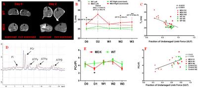
Mdx mice showed a significant increase in absolute T2 value compared to the WT group (figures A & B). Both groups (Mdx and WT) showed an acute increase in T2 in the exercised (right) leg at Day 2 following injury. However, the increase was more severe in the Mdx group and resolved by day 7 (W1). There also appeared to be a trend toward increasing T2 in both WT and Mdx exercised muscle at the end of study compared to the contralateral control leg (figure B). While a negative correlation exists between the T2 values and ULF acutely and chronically (figure C), there appeared to be a disconnect between T2 recovery and limb force recovery. PCr/Pi ratios showed no differences between groups at baseline (figure E), however, this ratio decreased in the Mdx group acutely upon exercise induced damage and resolved by day 7 (W1) similarly to the T2 values. In addition, there was a positive correlation observed between the PCr/Pi ratios and ULF (figure F).Conclusion:
Both T2 and energetic measurements in muscle showed similar profiles to the previously published data in mice of similar age (1); however, they provide an additional and profound acute indication of injury in the Mdx muscle injury mouse model. This acute exercise induced muscle damage biomarker assessment may provide a window of opportunity to test new therapies that target improving skeletal muscle pathophysiology.Acknowledgements
No acknowledgement found.References
Heier CR, Guerron AD, Korotcov A, et al. Non-invasive MRI and spectroscopy of mdx mice reveal temporal changes in dystrophic muscle imaging and in energy deficits. PLoS One 2014;9(11):e112477.Figures

(A) Representative MR T2-weighted
images acquired at baseline (Day 0) and on Day 2 after inducing the muscle
damage. The T2 values (B) showed a significant increase on day 2 (D2)
post injury, and a significant correlation (C) with the ULF. Representative 31P
MR spectra (D) showed a reduced PCr peak in the Mdx mouse (red) compared to the
WT mouse (blue) hind limb muscle. The PCr/Pi ratio (D) showed a significant
decrease at day 1 (D1) post injury compared to day 0 (D0) and day 7 (W1). The
PCr/Pi ratio significantly correlated with the ULF (E).