4975
Ultra-fast abdominal imaging with high parallel-imaging factors: Comparative study of a 60-channel receiver coil with the standard coil set-up1Radiology, University Hospital Tübingen, Tübingen, Germany, 2Radiology, University Hospital Tübingen, 3Siemens Healthineers
Synopsis
In this study, we evaluated a novel 60-channel coil setup for ultra-fast abdominal imaging using high PAT factors in a phantom, in healthy volunteers and in patients. We found that the 60-channel coil-setup is superior to a conventional 30-channel coil-setup yielding higher SNR and superior image quality and enabling ultra-fast image acquisition with diagnostic image quality.
Purpose
To assess the applicability and advantage of a novel 60-channel receiver coil setup in ultra-fast abdominal imaging with high parallel acquisition technique (PAT) factors with special regards to Signal-to-Noise-Ratio (SNR) and subjective image quality as compared to a standard 30-channel coil set up.Methods
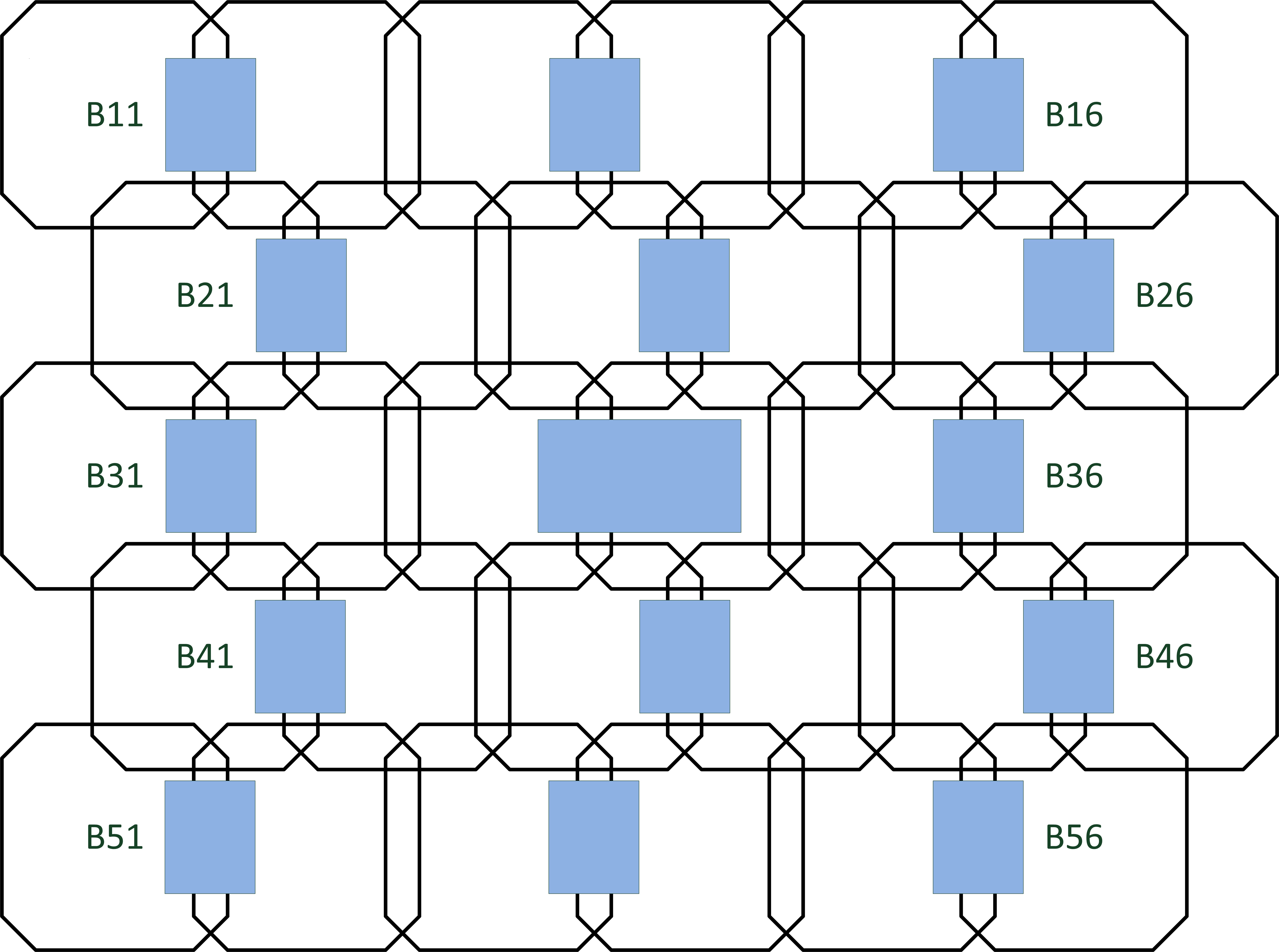
All imaging data were acquired on a 3T MR scanner (Magnetom Skyra, Siemens Healthcare, Erlangen, Germany). 3D T1 GRE imaging with different PAT factors (none, 2, 3, 2x2, 3x2, 2x3, 3x3) using “Controlled Aliasing in Parallel Imaging Results in Higher Acceleration” (CAIPIRINHA) (1) was performed in an oil phantom as well as in 5 healthy volunteers using a novel 60-channel (30-channel anterior and posterior) receiver coil setup (see Figure 1) in addition to a standard 30-channel (18-channel body and 12-channel spine coil)-setup. SNR was measured on phantom images using the subtraction method (2). Furthermore, overall image quality, image noise, artifacts, image quality of covered organs were assessed by two radiologists for all abovementioned sequenced on a 5-point Likert-scale (1=very poor, 2=poor, 3=moderate, 4=good, 5=excellent quality). In a further step, standard contrast-enhanced abdominal 3D T1 GRE imaging with CAIPIRINHA PAT factor 2 as well as accelerated imaging with CAIPIRINHA PAT factors 2x2 and 3x2 was performed in 17 patients who were randomly assigned into two groups (group A: 60-channel coil-setup, n=8; group B: 30-channel coil-setup, n=9). Image quality as described above was assessed and compared between the two patient groups.Results
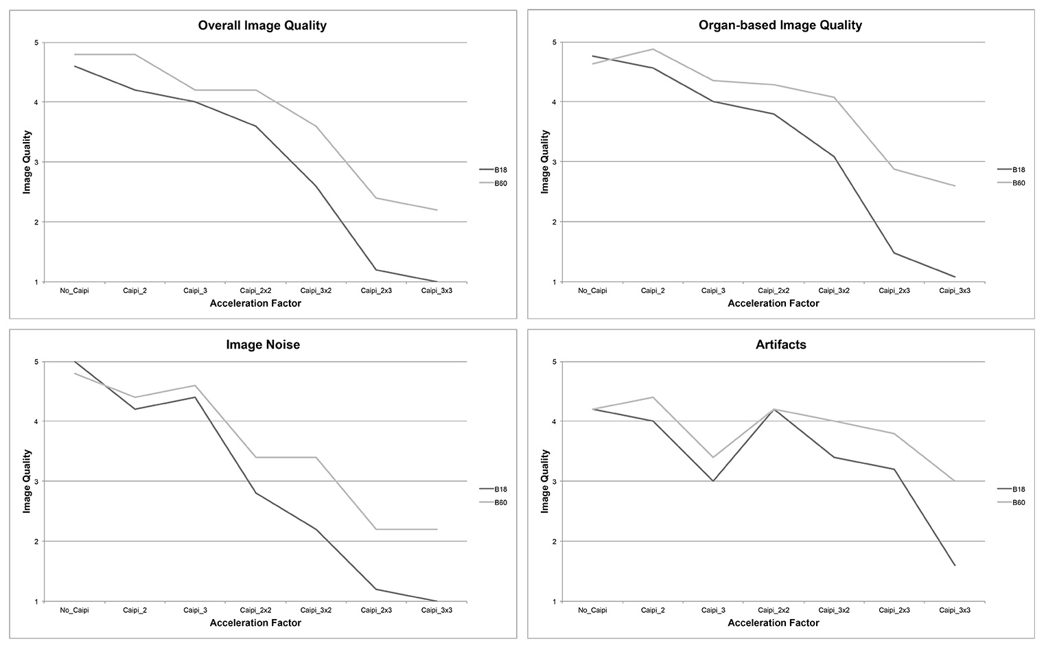
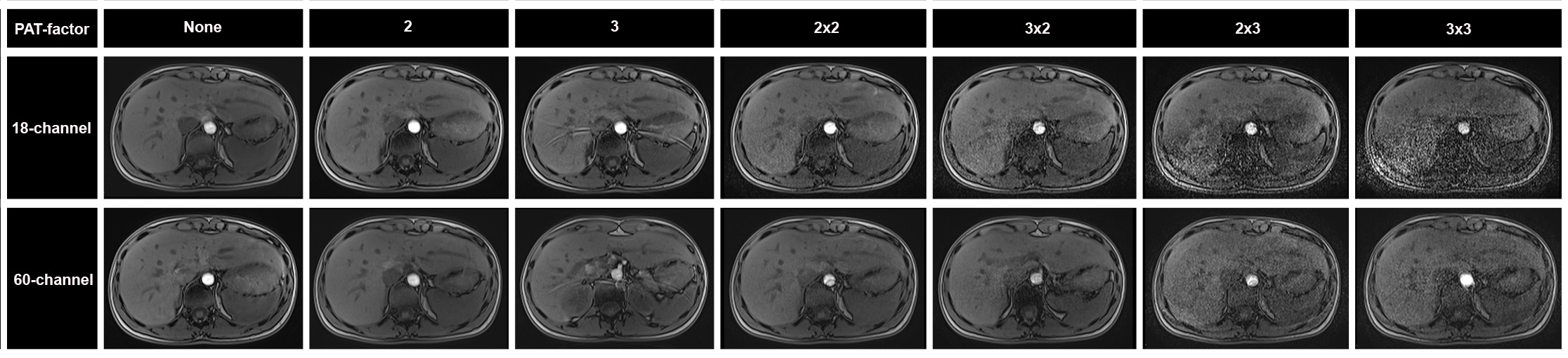
In comparison to the 30-channel coil, SNR gain for the 60-channel setup was observed for all acquisitions except for PAT=none. Relative SNR gain for the 60-channel coil ranged between 2.21% for PAT=2 and 22.69% for PAT=3x2. Comparison of image quality in healthy participants revealed significantly higher ratings of the 60-channel setup for all acquisitions (p≤.046), except for PAT=none (p≥.180), see Figures 2 and 3. In patients, similar results were observed with comparable image quality at lower PAT factors (PAT=2; p≥.069) and significantly superior image quality of the 60-channel setup for higher PAT factors (2x2 and 3x2; p≤.036).Discussion
The results of this study indicate that the use of a 60-channel setup increases SNR and image quality in abdominal 3D T1 GRE imaging. The benefit of the 60-channel coil-setup is mainly observed at higher PAT factors enabling ultra-fast imaging at CAIPIRINHA PAT factors up to 3x2.Conclusion
In comparison to a standard 30-channel coil-setup, the 60-channel coil-system improves image quality in abdominal 3D T1 GRE imaging and therefore enables ultra-fast imaging at high PAT factors with diagnostic image quality.Acknowledgements
NoneReferences
1. Breuer FA, Blaimer M, Heidemann RM et al. Controlled aliasing in parallel imaging results in higher acceleration (CAIPIRINHA) for multi-slice imaging. Magnetic resonance in medicine 2005; 53(3): 684-691.
2. Firbank MJ, Coulthard A, Harrison RM et al. A comparison of two methods for measuring the signal to noise ratio on MR images. Physics in medicine and biology 1999: 44(12): N261.
Figures