4974
Diffusion-weighted MR Enterography imaging of the ileocecal segment: optimizing b-value for visually differentiating inflammatory and neoplastic lesions1Department of Radiology, Tongji Hospital Tongji Medical College Huazhong University of Science and Technology, Wuhan, People's Republic of China
Synopsis
To evaluate the ability of conventional MR Enterography (MRE) including coronal and axial T1/T2 weighted imaging and Diffusion-weighted imaging with different b-values(b=400,600,800,1000,1200,1500,3000 sec/mm2)to visually illustrate inflammatory lesions and neoplastic lesions in the ileocecal region comparing with colonoscopy or surgical results.As a result,MRE and DWI were capable of revealing the lesions in the ileocecal segment. DWI was superior to detect lesions especially inflammations comparing with conventional MRE, and the optimal b value of DWI for MRE was 800 sec/mm2 at 3T. Hyperintensity of ileocecal lesion on DWI with high b(﹥1000 sec/mm2) value wes more favor for tumor-like lesion.
main abstract
Purpose: To evaluate the ability of conventional MR Enterography (MRE) including coronal and axial T1/T2 weighted imaging and Diffusion-weighted imaging with different b-values in diagnosis and differentiation of inflammatory lesions and neoplastic lesions in the ileocecal region comparing with colonoscopy or surgical results.
Materials and Methods: This study was approved by the institutional review board, and written informed consent was received before examination. Seventy-six patients suspected with ileocecal lesions were examined with conventional MRE and DWI with different b-values in the 3.0T MRI scanner (Discovery 750, GE Medical System, Milwaukee, USA) using a 32-channel torso phased-array coil. The parameters of MRE: (1) axial and coronal FIESTA with and without fat suppression [repetition time (TR), 3.2 ms; echo time (TE), 1.2 ms; matrix, 224 × 224; flip angle, 45°; slice thickness, 5mm; gap, 1mm];(2)axial and coronal T2-weighted single-shot fast spin echo with fat suppression (TR, based on the respiratory interval;TE, 68 ms; matrix, 256 × 256; slice thickness, 5mm; gap, 1 mm); (3) axial and coronal 3D fast field echo (LAVA) T1 sequence (TR, 3.82ms; TE, 1.69ms; matrix, 210×192; slice thickness,5mm no gap). The DWI parameters: TR, 3000ms; TE, 62ms, matrix, 160 × 128; slice thickness, 6mm; gap, 1 mm; the center location was in ileocecal region; the b=400, 600, 800, 1000, 1200, 1500, 3000sec/mm2. All the images were transmitted to PACS; two experienced radiologists independently evaluated the radiological signs of DWI hyperintensity with different b-values in the ileocecal segment; the presence and absence of DWI hyperintensity were rated positive and negative results. When meeting the different results, the two radiologists coordinated consistent results together. Within two weeks all of the patients were accepted with colonoscopy or surgery as gold standard.
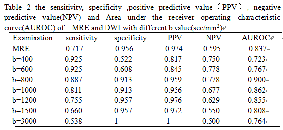
Results: By the gold standard, 19 of the patients were diagnosed with tumors, 34 were inflammations and 23 were normal. Forty-seven patients were found positive results in conventional MRE examination, the sensitivity, specificity ,positive predictive value(PPV), negative predictive value(NPV) were 0.758, 0.956, 0.979, 0.595.The positive results of DWI with different b-values were summarized with table 1. The sensitivity, specificity of different b values (b=400,600,800,1000,1200,1500,3000 sec/mm2) were 0.925, 0.925, 0.887, 0.811, 0.755, 0.660, 0.538; 0.522, 0.608, 0.913, 0.913, 0.957, 0.957, 1(table2).The results of two radiologists were with excellent consistency; the kappa values were 0.86 for conventional MRE and 0.64,0.72,0.82,0.95,0.98,0.95,0.95 for DWI with different b-values(b=400,600,800,1000,1200,1500,3000 sec/mm2).
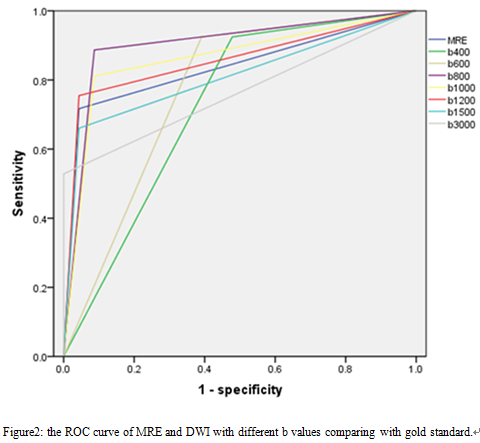
Discussion: DWI is a functional MR imaging technique that reflects the diffusion of water molecules in tissues [1]; the most critical parameter of DWI is the b values. The choice of optimal b values in the corresponding organization is conducive to lessons detection and identification. In the present study, it was found that DWI with b=800sec/mm2 had the appropriate sensitivity and specificity (table1 and figure2). The use of high b value DWI may be specific to hint tumor lesions[2];the signal intensity of inflammation lesions probably be suppressed (figure1) and we observed high ratio (60.7%,17/28) of tumor lesions in the positive results of DWI with high b value(b=3000sec/mm2).
Conclusion: MRE and DWI were capable of illustrating the lesions in the ileocecal segment. DWI was superior to detect lesions especially inflammations comparing with conventional MRE, and the optimal b value of DWI for MRE was 800 sec/mm2 at 3T. Hyperintensity of ileocecal lesion on DWI with high b(﹥1000 sec/mm2) value wes more favor for tumor-like lesion. For differential diagnosis, high b value DWI (up to 3000 sec/mm2) was recommended and a reliable noninvasive exam for patients suspected with ileocecal lesions.
Acknowledgements
The authors declare no relationships withany companies whose products or services may be related to the subjectmatter of the abstact.References
References:
[1]. Kim, K., et al., Diffusion-weighted MR Enterography for Evaluating Crohn's Disease. Inflammatory Bowel Diseases, 2015. 21(1): p. 101-109.
[2]. Fukukura, Y., et al., Diffusion-weighted MR imaging of the pancreas: optimizing b-value for visualization of pancreatic adenocarcinoma. European Radiology, 2016. 26(10): p. 3419-3427.
Figures
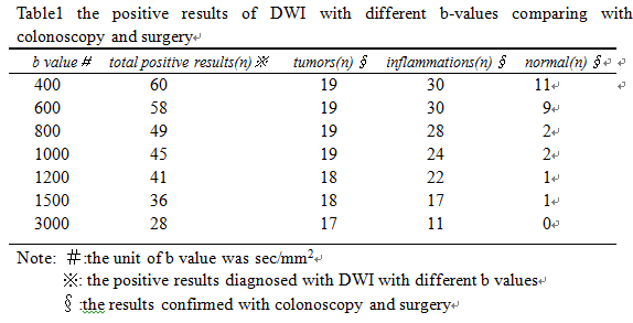
Table1 the positive results of DWI with different b-values comparing with colonoscopy and surgery