4971
Clinical application of 3D VIBECAIPI-DIXON for enhanced imaging of the small intestineYang Yu1, Lu Liang1, and Tao Jiang
1Beijing Chaoyang Hospital, Beijing,China, People's Republic of China
Synopsis
The abstract discussed the clinical application of a fast 3D VIBE sequence with Dixon fat saturation and CAIPIRINHA acceleration techniques (3D VIBECAIPI-DIXON) by compare to a standard 2D FLASH sequence with spectral fat saturation and conventional GRAPPA acceleration technique (2D FlashGRAPPA-fs) for enhanced imaging of the small intestine
Purpose
To compare a fast 3D VIBE sequence with Dixon fat saturation and CAIPIRINHA acceleration techniques (3D VIBECAIPI-DIXON) to a standard 2D FLASH sequence with spectral fat saturation and conventional GRAPPA acceleration technique (2D FlashGRAPPA-fs) for enhanced imaging of the small intestine.Methods and materials
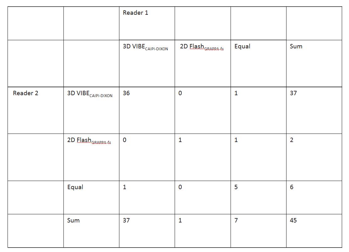
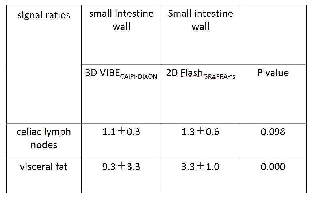
In this retrospective, 45 patients (18 female, 27 male) examined on a 18-channel 3.0-T MR system (Prisma, Siemens Healthcare Sector, Germany) were included; 3D VIBECAIPI-DIXON (TR/TE:4.21/1.35 ms; FOV:450mm; acquisition time:0.21min ) and 2D FlashGRAPPA-fs (TR/TE:221/2.46ms; FOV:450mm; acquisition time:0.53min) coronal sequences were performed in each subject in random order after the administration of an intravenous contrast agent. Two radiologists evaluated the images with regard to diagnostic preference; Semiquantitative signal ratios were calculated for the small intestine wall versus the visceral fat, and celiac lymph nodes. Signal ratio results were analyzed using a univariate analysis of variance.Results
3D VIBECAIPI-DIXON was preferred in 82.2% (both readers) and 2D FlashGRAPPA-fs in 2.2%/4.4% (reader 1/2) of cases with a kappa value of 0.779. The main reasons for this preference were homogenous fat saturation with 3D VIBECAIPI-DIXON and reduced motion artifacts due to a faster acquisition, leading to improved delineation of the small intestine wall. Signal ratios of small intestine wall to fat signal for 3D VIBECAIPI-DIXON (9.3±3.3) and 2D FlashGRAPPA-fs (3.3±1.0) were statistically different (P<0.05). However, no additional statistically significant differences in signal ratios were identified (range: 1.1±0.3 to 1.3±0.6; P>0.05).Conclusion
3D VIBECAIPI-DIXON can get small intestine imaging with a shorter time and improved fat suppression relative to conventional 2D FlashGRAPPA-fs; 3D VIBECAIPI-DIXON can help patients who can not hold breath for a while to acquire a better image.Acknowledgements
No acknowledgement found.References
No reference found.Figures
Table
1 Qualitative assessment by both readers
Table
2 Signal ratios between different portions of the small intestine wall and the
visceral fat, and celiac lymph nodes