4959
Differentiation of low- and high- grade hepatocellular carcinomas with texture features and a machine learning model in arterial phase of contrast-enhanced MR1Shenzhen Institutes of Advanced Technology, Chinese Academy of Sciences, Shenzhen, People's Republic of China, 2Department of Radiology, Guangdong General Hospital, Guangdong Academy of Medical Sciences
Synopsis
Texture has been a recognized feature for biological aggressiveness of hepatocellular carcinomas (HCCs). However, texture feature alone may not be optimal to characterize malignancy of HCC. Computer-aided techniques combined with multi-feature fusion may be a method of choice for the preoperative assessment of the aggressiveness of HCC. To this end, a computer-aided method in the combination with machine learning technique based on texture analysis for malignancy differentiation of HCCs was desmonrated and high classification performance(AUC>0.9) of the classifier was achieved to differentiate low- and high- grade of HCCs.
INTRODUCTION:
Clinical grading of the histological differentiation of Hepatocellular Carcinoma (HCC) is significant to establish a therapeutic strategy and to predict the prognosis 1,2. Computer-aided methods in the combination with machine learning techniques have recently shown a great potential in providing additional informationof the malignancy of HCC lesions3. In this study, we aimed to differentiate the malignancy of HCCs using texture analysis combined with a machine learning technique Support Vector Machine(SVM) based on the Gd-DTPA-enhanced MR images, and developed a classification model using feature selection of texture features in contrast enhanced MR images to accurately differentiate histological grading of HCCs.METHODS:
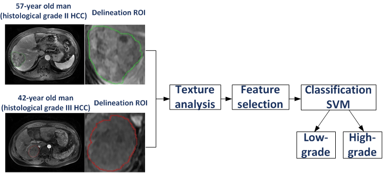
Fourty six subjects with pathologically proven HCCs who underwent Gd-DTPA-enhanced MRI exams from October 2011 to September 2015 were included and grouped into high differentiated group (Edmondson grade I and II) and low differentiated group (Edmondson grade III and IV). MR images were acquired with a 3.0T scanner (Signa Excite HD 3.0T, GE Healthcare, Milwaukee, WI, USA) and eight-channel phase-array coil using a breath hold Axial LAVA+C(liver acquisition with volume acceleration, LAVA) protocol. The framework of the proposed method for predicting grades of HCCs was illustrated in Figure 1. The region of interests (ROIs) was manually defined on the axial slice (arterial phase) with the largest tumor diameter by an experienced radiologist (10 years of experience in abdomen radiology). A total of 141 features were calculated for each ROI based on three kinds of texture analysis methods including histogram-based feature analysis, gray-level co-occurrence matrix (GLCM) and gray-level run-length (GLRL). Feature selection using the SVM-recursive feature elimination (SVM-RFE)4 algorithm was adopted to obtain a subset of most discriminative features that provided optimal performance. Support vector machine (LibSVM)5 was selected as the classifier to differentiate those lesions into high-grade or low-grade HCCs based on the selected features with optimal parameters determined by cross-validation. The four-fold cross-validation with 10 repetitions evaluation and the leave-one-out validation were used to assess the classifier. An alternative free-response receiver operating characteristic (ROC) analysis was performed to evaluate the classifier and the area under the standard ROC curves (AUC) was also calculated.RESULTS:
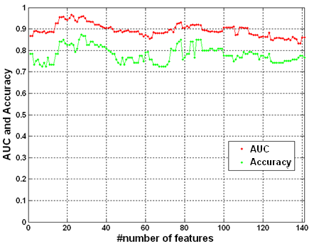
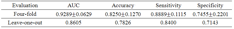
The peak AUC was achieved with 22 features selected by the SVM-RFE algorithm (Figure 2) with the biologic aggressiveness of HCCs reliably characterized by the most dominant texture features. The four-fold cross validation evaluation with 10 repetitions yielded AUC (0.9289±0.0629) with accuracy (0.8250±0.1270), sensitivity (0.8889±0.1115) and specificity (0.7455±0.2201) to differentiate low- and high-grade of HCCs (Table 1). The leave-one-out evaluation resulted in AUC (0.8605) with accuracy (0.7826), sensitivity (0.8400), and specificity (0.7143) (Table 1).DISCUSSION:
The proposed approach based on texture feature selection and SVM classification proved to be valid in differentiating low- and high- differentiated HCCs with high classification accuracy(AUC>0.9) (Table 1). The feature selection process based on the SVM-RFE algorithm resulted in higher classification performance compared with the case of using all the features (Figure 2). The top fifiteen to twenty features returned by the SVM-RFE algorithm might be appropriate to obtain high classification performance. In particular, Mean, Run percentage, Gray level nonuniformity and Run length nonuniformity were frequently top ranked features, which were consistant with the previous finding that Grey level nonuniformity and Mean could quantitatively characterize malignancy degrees of HCCs 6. The proposed method with machine learning technique provides a new perspective for automatically extracting the optimal features in terms of characterization of the biological aggressiveness of HCCs in addition to methods using texture feature alone.CONCLUSION:
The proposed approach using texture features and SVM is efficient in differentiating low- and high-grade HCCs based on the arterial phase of contrast-enhanced MR images, and showed the potential applicaition in the prodiction the overall malignancy of HCCs in the non-invasive manner that may facilitate better patient management in clinical practice.Acknowledgements
This research is partially supported by grants NSFC U1301258, NSFC 61302171, JCYJ20150630114942291 and No 2011S013.References
1. Parkin DM, Bray F, Ferlay J, Pisani P. Estimating the world cancer burden: Globocan2000. Int J Cancer 2001;94(2):153-156.
2. Llovet JM, Burroughs A, Bruix J. Hepatocellular carcinoma. Lancet 2003;362:1907–1917.
3, Larroza A, Moratal D, P.S Alexandra, et al. Support Vector Machine Classification of Brain Metastasis and Radiation Necrosis Based on Texture Analysis in MRI. J Magn Reson Imaging, 2015,42(5):1362-1368.
4. Ke Yan et al. Feature selection and analysis on correlated gas sensor data with recursive feature elimination, Sensors and Actuators B: Chemical, 2015;212:353-363.
5. www.csie.ntu.edu.tw/~cjlin/libsvm.
6. Wu Zhou et al. Malignancy Characterization of Hepatocellular Carcinomas Based on Texture Analysis of Contrast-Enhanced MR Images. J Magn Reson Imaging, 2016 (Online).
Figures