4955
Prostate cancer detection with multiparametric MRI based computer-aided diagnosis: which sequence is the dominant technique1Radiology, Peking University First Hospital, Beijing, People's Republic of China
Synopsis
Differ from PI-RADS v1, the updated PI-RADS v2 offers a decision process that puts the sequences as different role in scoring process and results in a final five-point score. However, the efficiency of each sequence in prostate cancer (PCa) detection in peripheral zone (PZ) and transition zone (TZ) is investigated by radiologists reading test preliminarily, which is highly depends on reader’s expertise and experience. This work applied a previous published machined learning model to investigate the weight of different sequences, including T2WI, DWI/ADC and DCE, in clinical significant PCa detection, and found that DWI/ADC performed the best both in PZ and TZ clinical significant PCa detection among these basic sequences which is recommended by PI-RADS v2
Introduction
PI-RADS v2 recommends1 that DWI/ADC and T2WI play more important roles in peripheral zone (PZ) and transition zone (TZ) clinical significant (CS) prostate cancer (PCa) detection, respectively.2,3 While DCE plays a minor role only in score 3 PZ lesions assessment.4 In this study, we validate a dedicated computer aided diagnosis (CAD) system to investigate which sequence is the dominant technique for CS PCa detection.Materials and Methods
Study Population:
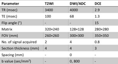
Our institution’s ethics review board approved this retrospective study. Between Dec. 2008 and Jan. 2010, 71 consecutive patients (mean age 68.3±7.7 years, range 40-82 years) who were suspicious with PCa in the prostate MR database were selected for the retrospective study. All enrolled patients underwent subsequent transrectal ultrasound (TURS) guided random systematic biopsy within 3 months and long-term follow-up (12 to 59 months, mean 32 months). All MR images of prostate multiparametric MRI (mpMRI) were carried out on a 3.0 Tesla MR scanner ( Signa TM; GE Medical Systems, Milwaukee, WI), using a abdominal coil. The imaging sequence parameters are summarized in Table 1.
Data Analysis:
The regions of interest (ROIs) with 6×6 pixels were manually drawn in the most suspected regions in PZ and TZ separately on T2WI by an experienced radiologist and a pathologist in consensus according to TRUS-guided biopsy. Biopsy-based definition of CS PCa was Gleason ≥ 3+4 or Gleason 6 with maximal cancer core length ≥ 4mm. The mpMRI features of each ROIs, including general features, gray-level histogram features and co-occurrence matrix (GLCM) features of T2WI and DWI/ADC, semi-quantitative features that calculated from DCE series were extracted. Then the image features of T2WI, DWI/ADC and DCE of PZ and TZ were used to be the inputs of the artificial neural network (ANN) classifier, respectively.
Statistical Analysis:
The effectiveness of each sequences for PZ and TZ CS PCa detection by application of the CAD system was evaluated by the receiver operating characteristic (ROC) analysis. During the training and evaluation process, ROIs in the PZ and TZ were analyzed separately. Leave-one-patient-out cross-validation method was used to separate the dataset into a training set and a test set. The correlation strength between the prediction of the CAD system and the PI-RADS V2 scores of T2WI and DWI/ADC were obtained by the Spearman correlation test. All statistical tests were two sided, and P < 0.01 was considered to indicate significant difference.
Results
238 ROIs selected from PZ
(92 PCa and 146 non-PCa), and 188 ROIs from TZ (52 PCa and 136 non-PCa) were
enrolled in this study. The proposed CAD system achieved AUC of 0.931±0.019 and 0.909±0.029 for PZ and TZ. The accuracy was 0.883 For PZ and 0.915 for TZ. The effectiveness of
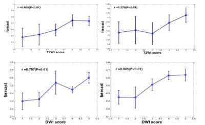
each sequences achieved by CAD system was shown in table 2. The Spearman
correlation coefficients between the prediction of CAD system and T2WI or
DWI/ADC scores were 0.600/0.787 (P<0.01) for PZ, and 0.379/0.805 (P<0.01)
for TZ (Fig 1).
Discussion/Conclusion
The CAD system based on mpMRI is effective in detection of CS PCa and the DWI/ADC sequence performs more effective than the other two sequences in identification of CS PCa both in PZ and TZ. Compare with the dominant role of T2WI sequence in identification of TZ CS PCa which improved by radiologists reading test,2 the poor correlation between CAD prediction and T2WI scores might indicate that T2WI interpretation involved more radiologists’ experience and expertise.Acknowledgements
References
1. Weinreb J C, Barentsz J O, Choyke P L, et al. PI-RADS Prostate Imaging - Reporting and Data System: 2015, Version 2 [J]. European urology, 2016, 69(1): 16-40.
2. Langer D L, van der Kwast T H, Evans A J, et al. Prostate cancer detection with multi-parametric MRI: logistic regression analysis of quantitative T2, diffusion-weighted imaging, and dynamic contrast-enhanced MRI [J]. Journal of magnetic resonance imaging : JMRI, 2009, 30(2): 327-334.
3. Hoeks C M, Hambrock T, Yakar D, et al. Transition zone prostate cancer: detection and localization with 3-T multiparametric MR imaging [J]. Radiology, 2013, 266(1): 207-217.
4. Oto A, Kayhan A, Jiang Y, et al. Prostate cancer: differentiation of central gland cancer from benign prostatic hyperplasia by using diffusion-weighted and dynamic contrast-enhanced MR imaging [J]. Radiology, 2010, 257(3): 715-723.
Figures


