4939
The detectability of mammary lesions dependent on the patients’ arm position and breathing style during a liver studyYasuo Takasu1,2, Yuko Shimada1, Tosiaki Miyati2, and Toshiki Shiozaki1
1Osaka Red Cross Hospital, Osaka, Japan, 2Division of Health Sciences, Graduate School of Medical Sciences, Kanazawa University., Kanazawa, Japan
Synopsis
This study aimed to evaluate whether the incidental finding of the mammary lesions was influenced by the patient’s arm position and respiration style during liver magnetic resonance imaging (MRI). If the mammary lesion was detected earlier, treatment could be performed earlier. Therefore, the finding of mammary lesion during liver studies was useful information. The incidental detection of mammary lesions was influenced by the patient’s arm position and breathing style. Unexpected mammary lesions could be detected when the arms were positioned at the sides of the body and the exhalation style was used during liver MRI.
Purpose
When magnetic resonance imaging (MRI) and computed tomography (CT) are planned, the target organs are included in the scan area. Sometimes, when these studies are performed, not only target lesions but also unexpected lesions are detected. Liver CT was routinely performed before liver MRI at Osaka Red Cross Hospital. We experienced that a mammary lesion was unexpectedly found during liver MRI, rather than during liver CT. This reason could be the different arm positions and respiration styles adopted during MRI and CT. A raised arm position was used during liver CT, because this position enables a decreased exposure dose [1] and increased image quality [2]. In contrast, the patients’ arms were at the sides of the body during liver MRI. This study aimed to evaluate whether the incidental finding of the mammary lesions was influenced by the patient’s arm position and respiration style during liver MRI and CT.Methods
The study included 473 consecutive female patients who underwent liver MRI with gadolinium contrast medium, and 2759 consecutive female patients who underwent liver CT with iodinated contrast medium, between May 2015 and April 2016. These studies were performed using a 1.5-T MRI unit and a 16- multidetector row CT. All patients who underwent liver MRI underwent liver CT. The detected mammary lesions were retrospectively extracted from diagnostic reports. Furthermore, the proportion of the breast included in the scan, for cases of incidental findings in the scan range, was evaluated using clinical images, with “bottom” defined as the edge of the foot direction of the breast and “top” as the edge of the head direction of the breast. (Fig. 1) We evaluated the proportion of the breast from “bottom” to the upper slice of liver MRI and CT.Result
No mammary lesions were found during liver CT. Seven mammary lesions were found during liver MRI. On the MRI images, the proportion of the breast within the scan range was as follows: whole breast for four cases, from bottom to between the nipple and the top for two cases, and from the bottom to the nipple for one caseDiscussion
The cases in which the mammary lesion was found during liver MRI but not during liver CT were influenced by the arm positon and respiration style. The raised arm position was useful during liver CT [1, 2]. However, the raised arm position was disadvantageous during liver MRI because the patient could become tired during the long examination term. Undue stress could cause the patient to move, leading to a poor study. In liver MRI, the arm position can be freely changed (i.e., the arms are positioned at the sides of the body) as long as there are no artifacts. When the arms were at the sides of the body, the patient could be more relaxed, with reduced mental stress during the examination; therefore, we believed that the arm position at the sides of the body was optimal. We considered that the reason for the detection of mammary lesions during MRI instead of CT was the arm position. When the arms were raised up over the head, the pectoralis muscle moved upward [3], so that the breast was outside of the scanned area. Furthermore, during the scan, the respiration style during liver MRI differed from that during liver CT. The liver position was changed by the movement of the diaphragm because of respiration [4]. When the exhalation style was used during liver MRI, the diaphragm and liver were moved up toward the head; therefore, we considered that the breast was more likely to be moved into the scan area. If the mammary lesion was detected earlier, treatment could be performed earlier and the quality of life would be influenced. Therefore, the finding of mammary lesion during liver studies was useful information. Therefore, with all other things being equal, if the same liver MRI were to be performed, the positioning of the arms at the sides of the body and the exhalation style would be preferable because of the higher probability of detecting an unexpected mammary lesions during liver MRI compared with that using the raised arm position and inhalation style.Conclusion
The incidental detection of mammary lesions was influenced by the patient’s arm position and breathing style during liver MRI and CT. Unexpected mammary lesions could be detected when the arms were positioned at the sides of the body and the exhalation style was used during liver MRI.Acknowledgements
No acknowledgement found.References
[1] Brink M, de Lange F, Oostveen LJ, et al. Arm raising at exposure-controlled multidetector trauma CT of thoracoabdominal region: higher image quality, lower radiation dose. Radiology 2008; 249: 661-670. [2] Karlo C, Gnannt R, Frauenfelder T, at al. Whole-body CT in polytrauma patients: effect of arm positioning on thoracic and abdominal image quality. Emerg Radiol 2011; 18: 285-293. [3] Yeh ED, Georgian-Smith D, Raza S, Bussolari L, Pawlisz-Hoff J, Birdwell RL. Positioning in breast MR imaging to optimize image quality. Radiographics. 2014 Jan-Feb;34(1):E1-17. [4] Langen KM, Jones DT. Organ motion and its management. Int J Radiat Oncol Biol Phys. 2001;50:265–278.Figures
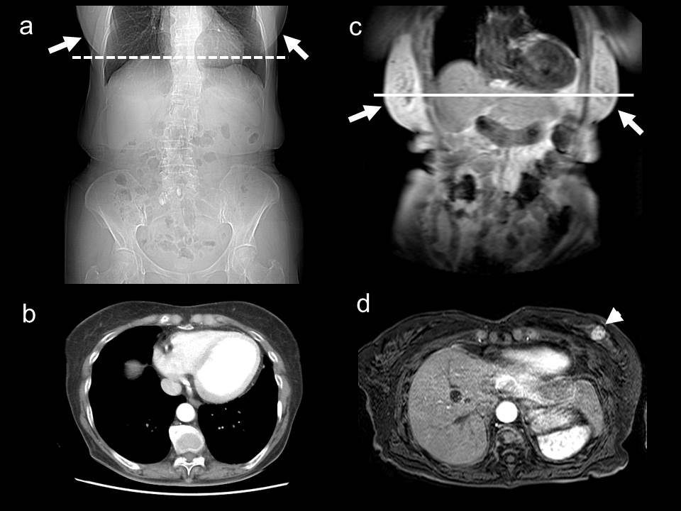
76-year-old woman with multiple hemangioma and hepatitis B
The scout view of
liver CT when the raised arm position and inhalation style were used (a), the
breast (arrow) was not found at the most upper slice (dotted line in a) (b).
The survey image of liver MRI when
the patients’ arms were at the sides of
the body and exhalation style was used (c), the breast (arrow) was
included with in the scan range; therefore, the breast tumor (arrow head) was
found (line in c) (d).
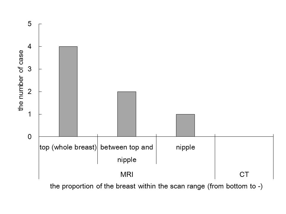
The number of case
for the proportion of the breast within the scan range
On the MRI images, the proportion of the
breast within the scan range in seven cases was as follows: whole breast (from
bottom to top) for four cases, from bottom to between the nipple and the top
for two cases, and from the bottom to the nipple for one case. No mammary lesions were
found during liver CT.