4936
Correction of image distortion and gradient nonlinearity in DTI of breast cancer1Radiology and Biomedical Imaging, University of California San Francisco, San Francisco, CA, United States, 2GE Global Research, Niskayuna, NY
Synopsis
The individual and combined effects of correction for susceptibility-induced distortion, distortion due to eddy currents, and bias from gradient non-linearity on breast DTI metrics were evaluated. Using an ice-water phantom we found that the correction of gradient nonlinearity resulted in strong bias reduction, while the distortion correction provided further reduction of bias and variance. The effects of these corrections were quantified in 12 subjects with malignant breast tumors and found to parallel the effects measured in the phantom.
Introduction:
In studies of breast cancer, DWI increased diagnostic accuracy and showed promise as a biomarker of early treatment response1. However, one limitation of breast DWI is that standard echo-planar imaging (EPI) suffers from geometric distortions resulting from the susceptibility-induced variation in the B0 field and eddy-currents from diffusion-encoding gradients. These distortion effects occur in addition to gradient non-linearity (GN) effects, which have been shown to affect the quantitative accuracy of breast DWI2. A robust method for correcting geometric distortions in the brain utilizes an extra reverse phase gradient (RPG) acquisition to estimate the B0 map3, and has also been recently applied to breast DWI4. However, there is little information about the effects of RPG based correction algorithms on the quantification of diffusion metrics in breast cancer. This work evaluates the individual and combined effects of correction for susceptibility-induced distortion, distortion due to eddy currents, and bias from GN on diffusion tensor imaging (DTI) metrics measured in a phantom and in malignant breast tumors.
Methods:
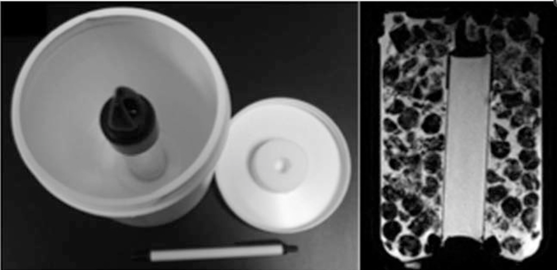
Diffusion tensor imaging data were acquired from bilateral ice-water phantoms with known ADC (1.1x10⁻³ mm2/sec) and FA (~0.0) values5 (Figure 1). In vivo DTI was acquired in 12 patients with biopsy-confirmed, locally-advanced breast cancer enrolled in an institutional review board-approved, HIPAA compliant, clinical trial. All patients gave informed consent and DTI data were acquired prior to initiation of treatment. Bilateral axial MRI were acquired on a 1.5T whole body scanner (GE Healthcare, Waukesha, WI) using an 8-channel breast coil (Invivo, Gainesville, FL). A standard DTI acquisition was performed (6 directions, b=0,600 s/mm2) along with a corresponding reversed-polarity-gradient (RPG) acquisition (1 direction, b=0, 600 s/mm2). Parametric ADC and FA maps were calculated from DTI data. Three corrections were evaluated: gradient nonlinearity correction (GNC)6, EPI distortion, and eddy-current (EC) effects. The EPI effects (phase-encoding distortion from inherent B0 susceptibility) were corrected using the RPG algorithm3. Eddy-current effects (phase-encoding distortion) were corrected using image registration, registering the diffusion images to the b=0 image assuming a cubic polynomial transformation model7. In the phantom, three axial slices were selected for analysis and trapezoidal ROIs of at least 1000mm2 area were drawn on the central water vials. The ROIs for non-RPG-corrected and RPG-corrected images were drawn separately, but keeping the ROI areas within 15%. For patient scans, one ROI was defined on the uncorrected DTI slice estimated to contain the largest tumor area. These ROIs were then mapped to the corresponding slice on the corrected ADC and FA maps; minor adjustments were made if needed. Differences between uncorrected and corrected ADC and FA were evaluated using a paired t-test, P<0.05 considered significant.
Results:
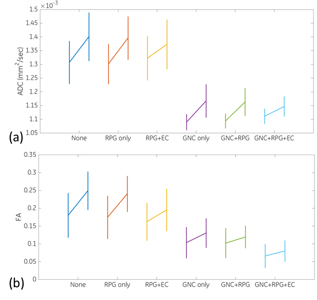
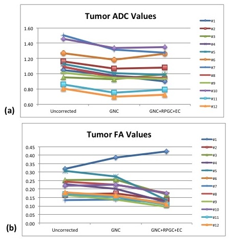
Figure 2 shows uncorrected and corrected DTI parameter maps of the two ice-water phantoms, with corresponding plots of ADC and FA values in Figure 3, demonstrating the dominant effect GNC has in reducing the bias of ADC and FA relative to the actual values of 1.1x10⁻³ mm2/s and 0. The addition of eddy current and RPG correction to GNC resulted in further improvement in the accuracy of the phantom ADC and FA measurements, with a stronger effect seen for FA. Figure 4 shows the effects of GNC only and GNC+RPG+EC corrections on mean tumor ADC and FA measurements from the patients. The GNC correction reduced tumor ADC (-9.35%), with a significant difference between pre- and post correction values (p<0.001), but it had no significant effect on FA (p=0.5). Addition of RPG and EC corrections to GNC did not significantly alter tumor ADC (p=0.79), but did significantly decrease FA (p=0.002), with an average change of -23.5%.
Discussion
Previous work has shown that gradient non-linearity can result in variation of tumor ADC and FA values, and that these variations can be corrected. The changes in tumor FA and ADC after GNC correction measured in this work were consistent with previous findings2 and analogous to the phantom measurements. The results of this work show that RPG and EC correction, when applied in addition to GNC, yield further improvement in accuracy of ADC and FA measurements in a phantom. The addition of RPG and EC correction to GNC in in vivo DTI measurements of FA showed a similar effect to the phantom, however it had no significant effect on ADC.
Conclusion:
Addition of RPG and EC distortion correction demonstrates measurable improvement in ADC and FA values measured in a phantom with an analogous effect on breast tumor ADC and FA values measured in patients.
Acknowledgements
The authors acknowledge useful discussion with Ileana Hancu and Jonathan Sperl, National Institutes of Health Grants U01CA151235 and R01CA190299, and Susan G Komen Grant SAC110017References
1. Pickles MD et al, Magn Reson Imaging 2006; 24(7):843-7.
2. Newitt DC et al, JMRI 2015; 42(4)908-19.
3. Holland D et al, Neuroimage 2010; 50(1):175-183.
4. Teruel J et al, Magn Reson Med 2015; 74:1138–1144.
5. Malyarenko D et al, JMRI 2013; 37:1238–1246.
6. Tan ET et al, JMRI 2013; 38(2)448-53.
7. Klein S et al, IEEE TMI 2010; 29(1):196-205.
Figures
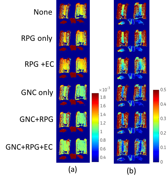
DTI parameter maps of the bilateral ice water phantoms showing the effects of different combinations of image corrections on (a) ADC and (b) FA values. The addition of the corrections bring the ADC value of the central water compartment closer to the expected value of 1.1x10⁻³ mm2/sec and the FA values also decreases toward the minimum of zero expected for an isotropic medium.