4923
High Resolution Breast Diffusion Weighted Imaging Using 2-D Navigated Multishot SENSE EPI with Image Reconstruction using Image-Space Sampling Function (IRIS) at 3 T1Radiology, University of Washington, Seattle, WA, United States, 2Body/Oncology, Philips Healthcare, Best, Netherlands, 3Radiology, Seattle Cancer Care Alliance, Seattle, WA, United States
Synopsis
Conventional diffusion weighted (DW) MRI relies on a single-shot (SS) echo planar imaging (EPI) acquisition, which suffers from limited spatial resolution and detrimental geometric distortions. Multishot (MS) EPI techniques hold potential to improve the image quality and spatial resolution of DW MRI. We tested the feasibility and performance for breast imaging of a recently developed DW MS-EPI sequence incorporating a novel IRIS image reconstruction approach. Our initial results demonstrate this DW MS-EPI technique to provide robust high resolution breast DW images, with good image quality and reduced geometric distortion compared to conventional DW SS-EPI.
Purpose
Diffusion-weighted (DW) MRI holds potential to improve the detection and biological characterization of breast cancer and is increasingly being incorporated into breast MRI protocols [1]. Conventional DW MRI sequences utilize a single-shot echo planar imaging (SS-EPI) readout to achieve fast motion-freezing image acquisition with high signal-to-noise ratio (SNR). However, the long readout echo train of SS-EPI renders it very sensitive to off-resonance effects due to magnetic field inhomogeneities, resulting in geometric distortions and limiting resolution, particularly at higher magnetic field field strengths [2]. These issues are further magnified for breast DWI due to the challenges of off-isocenter imaging and air-tissue interfaces, and can reduce accuracy and hinder direct comparison of DW MRI data with other imaging sequences. Alternatively, a multishot (MS) EPI approach divides the imaging phase encodes into multiple TR intervals, thus decreasing echo train length and resulting susceptibility-related distortions. However, MS-EPI requires complex motion-induced phase correction using navigator echoes or other methods to avoid ghost artifacts [3], which is technically demanding and has not been widely used for breast imaging. A new approach was recently introduced to perform DW interleaved 2-D navigated MS-EPI acquisitions with SENSE acceleration incorporating an efficient image reconstruction method that is less computationally and technically demanding than prior techniques [4]. We therefore aimed to test the feasibility and performance of this novel 2-D navigated MS-EPI acquisition for achieving high resolution breast DW imaging with low geometric distortion.Materials and Methods
In this IRB-approved prospective study, 17 women undergoing clinical breast MRI examinations consented to undergo both MS and conventional DW SS-EPI scans. MR imaging was performed on a 3T Philips MRI scanner with a 16 channel breast coil (Mammotrak, Philips). The two DW EPI sequences were performed with matching spatial resolution, slice positioning, and diffusion-encoding parameters. Both sequences used spin-echo diffusion preparation and spectral selection attenuated inversion recovery (SPAIR) fat suppression, SENSE=3, FOV=36x36cm, matrix=300x300, voxel size=1.2x1.2x4mm. Diffusion gradients were applied in three directions, with b-values of 0, 800 s/mm2. In both cases, the phase direction was anterior-posterior.
DW SS-EPI was performed with TR=6430ms, TE=66ms, averages=3, Δ=32ms, δ=13.8ms, scan time=1:36min. DW MS-EPI was performed with a dual spin-echo pulse sequence where the image-echo was followed by a navigator-echo with a 180° refocusing pulse between the two echoes, along with an efficient reconstruction method, called Image Reconstruction using Image-Space Sampling function (IRIS) [4]. DW MS-EPI imaging parameters were TR=6925ms, TE (image echo)=58ms, TE (navigator echo)=120ms, EPI shots=2, averages=1, Δ=28.9ms, δ=16.8ms, scan time=2:53min.
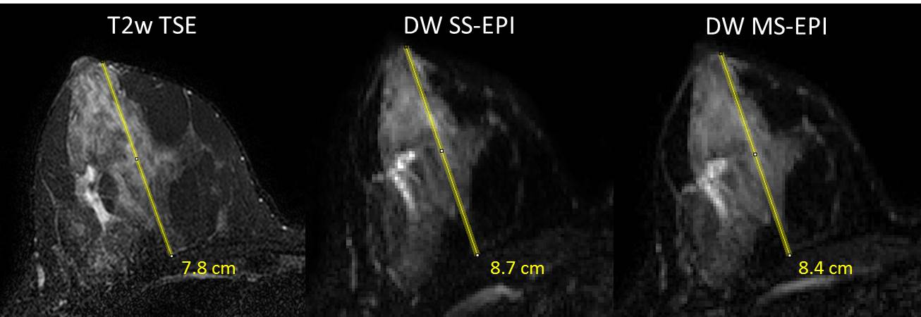
A T2-weighted turbo spin echo sequence (TSE) was acquired during the same examination and used as the reference standard for anatomical measures. Imaging parameters for the T2-weighted TSE images were TR=5000ms, TE=60ms, SENSE=3.1, FOV=22x33cm, matrix=275x412, voxel size=0.8x0.8x1.3mm, scan time 5:15min. DW images from both techniques were qualitatively and quantitatively compared for each subject. To assess spatial distortion, the anterior-posterior distance from nipple to chest wall was measured for both right and left breasts on DW SS-EPI and DW MS-EPI b=0 s/mm2 images and on T2-weighted TSE reference images, Figure 1. Absolute differences in nipple to chest wall distances (vs. T2-weighted TSE anatomic images) and differences in tissue ADC measures between DW SS-EPI and MS-EPI were evaluated by Wilcoxon signed rank test.
Results
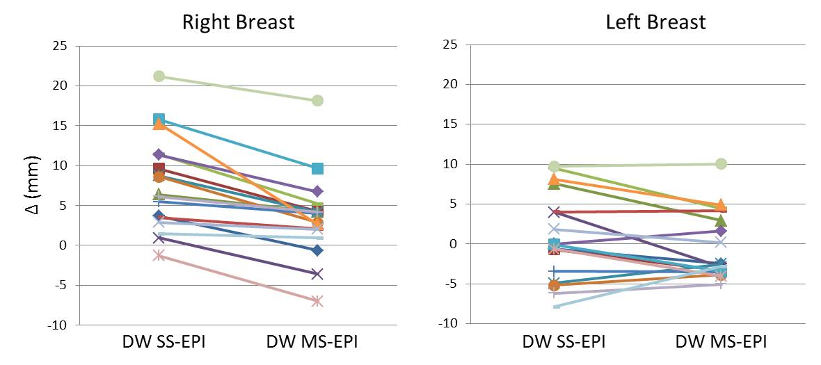
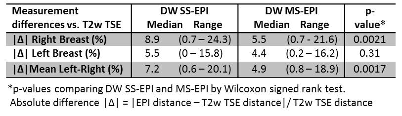
Overall, the DW MS-EPI sequence performed very well for breast imaging with no apparent ghosting observed and good SNR across all cases. DW MS-EPI images were visually observed to be similar to slightly superior in quality when compared to conventional SS DW EPI images for each subject in terms of off-resonance and blurring artifacts. Both DW EPI sequences exhibited differences in nipple-to-chest wall measures versus the T2-weighted TSE measures, which were significant (p<0.05) for right breast but not for left breast. However, the absolute differences in nipple-to-chest wall distances vs. T2w TSE were significantly less for DW MS-EPI compared to DW SS-EPI, as demonstrated in Figure 2 and Table 1.Conclusion
Our study is the first to investigate the use of a new 2D navigated DW MS-EPI approach for application in breast imaging. The novel DW MS-EPI sequence incorporates an efficient IRIS reconstruction that is less computationally and technically demanding than prior techniques, which may facilitate clinical implementation. Our findings demonstrate the technique enables robust high resolution breast DW MR imaging with reduced geometric distortion compared to conventional DW SS-EPI. Further work will investigate the optimization of this DW MS-EPI technique to achieve even higher spatial resolution breast imaging and improve lesion characterization.Acknowledgements
This research was supported by National Institutes of Health Grant R01CA151326 and a pilot grant from Earlier.Org - Friends for an Earlier Breast Cancer Test.References
1. Partridge SC, Nissan N, Rahbar H, Kitsch AE, Sigmund EE. Diffusion-weighted breast MRI: Clinical applications and emerging techniques. J Magn Reson Imaging. 2016. Epub 2016/10/01.
2. Le Bihan D, Poupon C, Amadon A, Lethimonnier F. Artifacts and pitfalls in diffusion MRI. J Magn Reson Imaging. 2006;24(3):478-88.
3. Bammer R, Holdsworth SJ, Veldhuis WB, Skare ST. New methods in diffusion-weighted and diffusion tensor imaging. Magnetic resonance imaging clinics of North America. 2009;17(2):175-204.
4. Jeong HK, Gore JC, Anderson AW. High-resolution human diffusion tensor imaging using 2-D navigated multishot SENSE EPI at 7 T. Magnetic resonance in medicine. 2013;69(3):793-802.
Figures