4918
Correlation of dedicated breast PET and dynamic contrast MRI: Appearance of breast background parenchyma and breast cancers1Radiology, Rakuwakai Otowa Hospital, Kyoto, Japan, 2Radiology, Stanford University School of Medicine, Stanford, CA, United States, 3Diagnostic Imaging and Nuclear Medicine, Kyoto University Hospital, Kyoto, Japan
Synopsis
A recently developed ring-shaped PET scanner dedicated for breast (dbPET) provides high-resolution 3D images of a breast. We investigated the correlation between dbPET with 18F-fluorodeoxyglucose and dynamic contrast MRI (DCE-MRI) findings in respect to breast background parenchyma and mass-forming breast cancers. Background parenchymal uptake on dbPET was not associated with background parenchymal enhancement on DCE-MRI. Tumor appearance on dbPET was similar to that on DCE-MRI in majority of cases, suggesting improved spatial resolution of dbPET as well as its feasibility for the use of the combined image analysis with DCE-MRI aiming at functional and structural assessment of primary breast tumors.
Purpose
A recently developed dedicated breast PET with a ring-shaped scanner provides high-resolution 3D PET images of a breast1. The aim of this study was to investigate the correlation of imaging findings between dbPET and breast dynamic contrast MRI (DCE-MRI) in respect to breast background parenchyma and mass-forming breast cancers.
Patients and Methods
A total 35 breasts of 35 female patients (32-80 yrs) with invasive breast cancer were retrospectively analyzed in this study. All patients underwent both 18F-fluorodeoxyglucose (FDG) dbPET and DCE-MRI in the prone position before treatment, and had an index tumor that appeared as a mass of 1 cm or larger on DCE-MRI. Four readers (2 breast radiologists and 2 nuclear medicine physicians) independently evaluated dbPET and DCE-MRI images of unilateral affected breasts for background parenchyma (amount and uptake grade of FDG-avid breast tissue on dbPET, amount of breast tissue on T1-weighted image, background parenchymal enhancement [BPE], tumor conspicuity to background) and index tumor appearance (shape, margin, internal pattern). All data were treated as bivariate data for analysis, and final categories were determined on all readers’ judgements by majority rule. Amount and uptake grade of FDG-avid background breast parenchyma on dbPET were compared with breast tissue amount on T1-weight image and BPE on early-phase DCE-MRI using Fisher’s exact test. McNemar test was performed to identify a difference between dbPET and DCE-MRI for each category of tumor appearance.Results
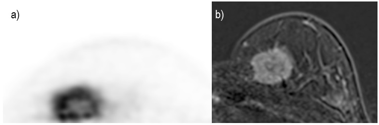
Amount of FDG-avid breast tissue on dbPET was large (occupying 50% or more of a breast) in 46%, and was significantly correlated with breast tissue amount on T1-weighted image (p<0.001) but not with BPE. Moderate to marked background parenchymal uptake was seen in 26% on dbPET, and was not associated with breast tissue amount on T1-weighted image nor BPE. Tumor conspicuity to background was good to excellent in all cases in both dbPET and DCE-MRI. In both modalities, majority of tumors had irregular shape (dbPET vs. early-phase DCE-MRI = 51% vs. 77%, p=0.066), irregular margins (66% vs. 80%, p<0.005), and heterogeneous internal uptake (77% vs. 97%, p<0.001). Rim uptake was observed in 29% of cases on dbPET, while rim enhancement was seen in 14% on early-phase DCE-MRI (p=0.131). In the side-by-side comparison, the similarity of index tumor appearance between dbPET and early-phase DCE-MRI was identified in 77% in shape, 60% in margin, and 51% in internal pattern (Figure).Discussion
While breast tissue amount on T1-weighted image was correlated with amount of FDG-avid parenchyma, our study showed that neither amount nor uptake grade of FDG-avid breast tissue on dbPET was associated with BPE on DCE-MRI, indicating a different tendency may exist between dbPET and MRI in the physiological parenchymal accumulation of agents. As to index tumor appearance, dbPET demonstrated similar morphological features to DCE-MRI in the majority of cases, suggesting that dbPET successfully provides high-resolution images up to a level relatively close to MRI, which has been difficult with low-resolution conventional whole body PET. Rim appearance appeared slightly easier to be identified with dbPET than DCE-MRI, although there was no significant difference.Conclusions
Background parenchymal uptake on dbPET was not associated with BPE on DCE-MRI. The frequent similarity of tumor appearance on dbPET to MRI suggests the improved capability of dbPET to visualize the detailed structures of breast cancers, and that dbPET images are feasible to be fused with MRI for the combined image analysis of dbPET and MRI possibly leading to functional and structural assessment of primary breast tumors.Acknowledgements
No acknowledgement found.References
1. Miyake KK, Matsumoto K, Inoue M, et al. Performance Evaluation of a New Dedicated Breast PET Scanner Using NEMA NU4-2008 Standards. J Nucl Med. 2014;55(7):1198-203.Figures