4914
Localized spectroscopy of hyperpolarized xenon-129 dissolved in the human head with a dedicated receiver array1University Of Sheffield, Sheffield, United Kingdom, 2GE Healthcare Inc, Aurora, OH, United States
Synopsis
This study investigates the spectral signal of hyperpolarized 129Xe dissolved in the human head in vivo using a dedicated radiofrequency receiver coil array. With a 2.5-fold higher signal to noise ratio of the array compared to a conventional transmit-receive radiofrequency coil, we detected 8 spectral peaks compared to 5 peaks reported in an earlier study. From this, we postulate an individual assignment of spectral peaks for hyperpolarized 129Xe dissolved in white matter and soft cartilaginous tissue, which were previously undistinguishable.
Purpose
129Xe is soluble in blood, can cross the blood-brain barrier and exhibits distinct resonant peaks when dissolved in the different tissues of the human head, making 129Xe MR spectroscopy a potentially powerful tool for assessment of cerebral perfusion. Spectroscopic studies of hyperpolarized (HP) 129Xe dissolved in the human head in vivo have to date used transmit-receive radiofrequency (RF) coils, which have enabled the detection of between 1 and 5 spectral peaks1-3. The purpose of this study was to enhance the signal to noise ratio (SNR) of HP 129Xe in the human head at 1.5 T through the design and build of a RF receiver coil array, which should not only improve SNR but also provide regional information based on the coils sensitivity profiles.Methods
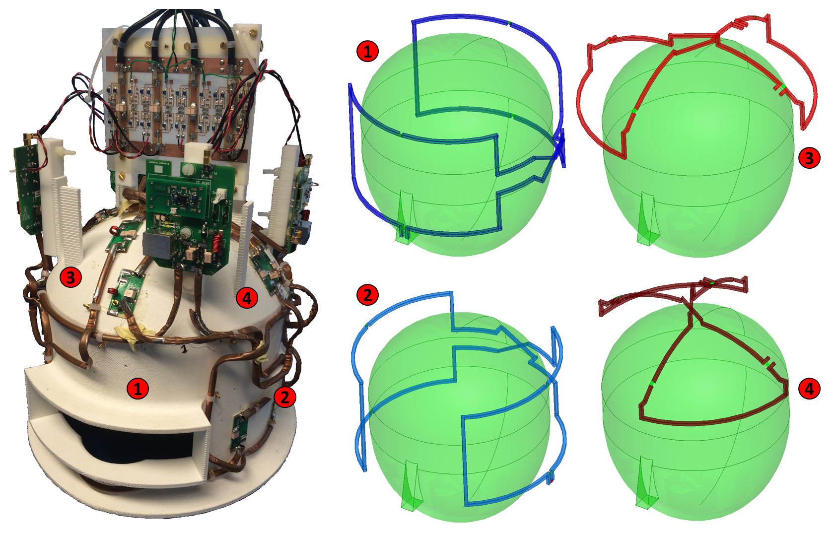
A 4 channel RF receiver coil array was developed in house with 0.3 mm thick hollow copper tube (CW008A) conducting elements on a 3D printed ABSplus mechanical former, as shown in Figure 1. Elements 1-2 cover the whole of the brain/upper-head and elements 3-4 cover the top of the brain/head, each element being a volume figure-of-eight. RF transmission was achieved with a low-pass 8 rung volume birdcage resonator built in house.
129Xe was hyperpolarized to ~ 20 % using a spin-exchange optical pumping polarizer4. Spectra were acquired from the heads of 3 healthy male volunteers (aged 33, 30 and 34 years) following inhalation of a xenon gas dose of 800 to 1000 mL, on a 1.5 T MR system. The spectral acquisition parameters were: hard RF pulse; pulse duration = 500 µs; receive bandwidth = 135 ppm; spectral resolution = 0.033 ppm; centre frequency = 198 ppm; flip angle = 45° and repetition time = 2 seconds. 10 spectra were acquired in a single breath-hold and averaged.
Results
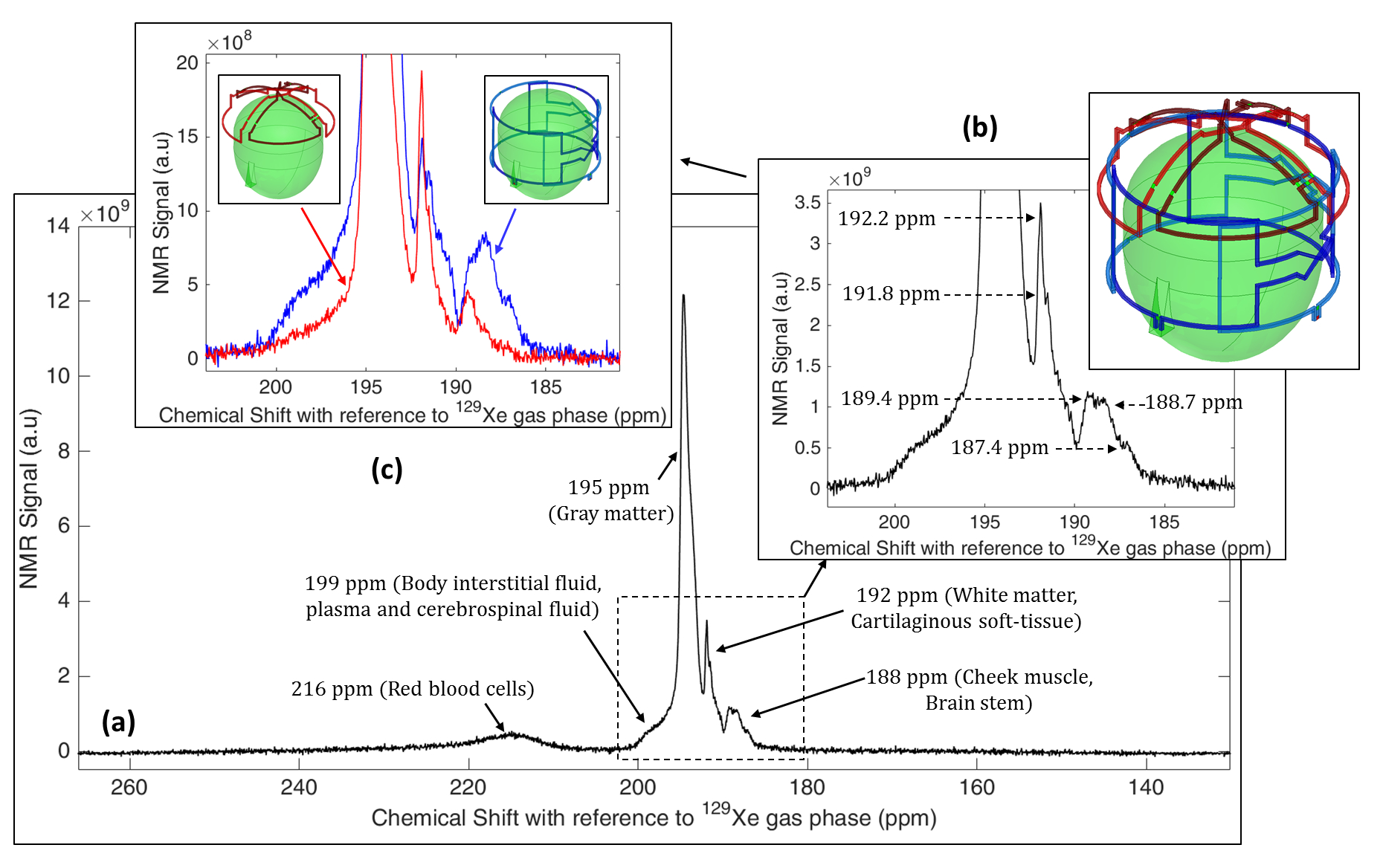
The unloaded and loaded quality factor (Q) of the RF array was 330 and 120, respectively. Isolation between the elements was < -18 dB. A typical averaged spectrum of HP 129Xe dissolved in the human head is shown in Figure 2(a). As seen in Figure 2(a) and (b), 8 peaks were detected at chemical shifts of 187.4 ppm, 188.7 ppm, 189.4 ppm, 191.8 ppm, 192.2 ppm, 195 ppm, 199 ppm and 216 ppm with respect to 129Xe in the gas phase. When the spectra from elements 1 and 2 (whole brain/upper-head) were analysed separately from 3 and 4 (top brain/head), an absence of the peaks at 187.4 ppm, 188.7 ppm and 191.8 ppm in the top of the head was observed (Figure 2(c)).
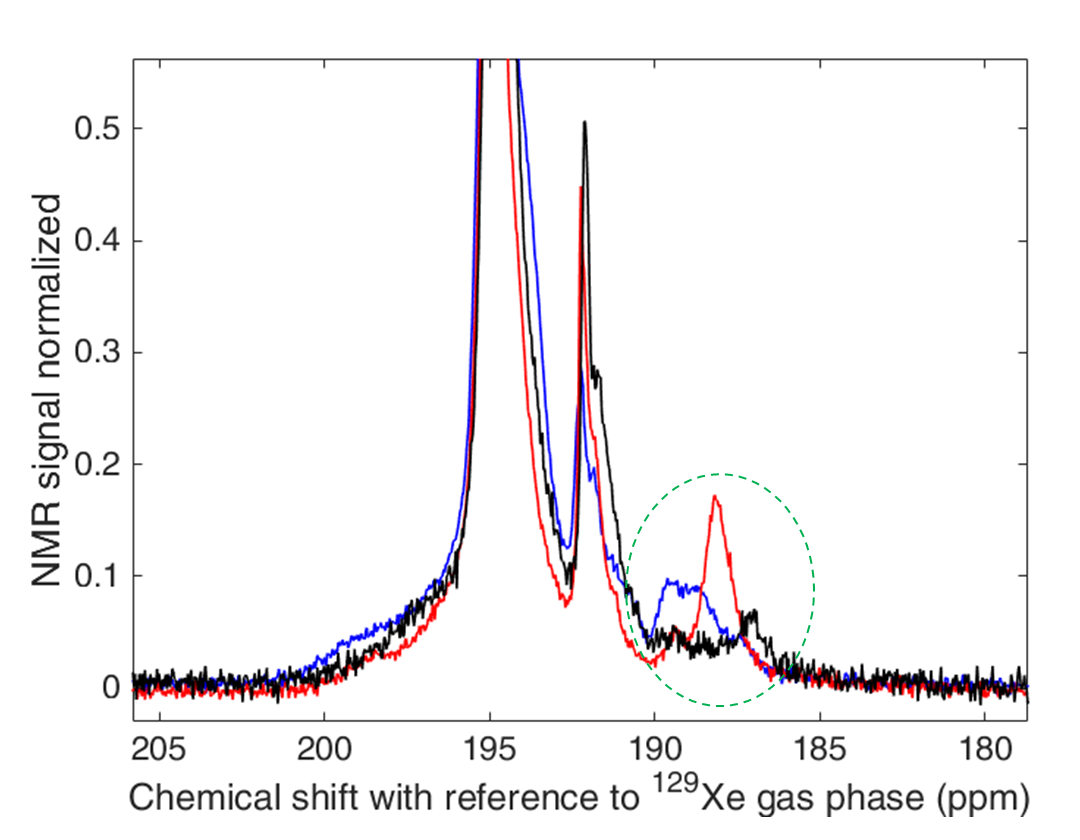
In the chemical shift range of 185-190 ppm, spectra from three different healthy volunteers exhibited significant differences in appearance of spectral peaks, such as presence / absence and their relative magnitude, as seen in Figure 3.
Discussion
An earlier study of human brain 129Xe spectroscopy and chemical shift imaging showed two sources (discrete chemical environments) contributing to a single spectral peak at 192 ppm; one source being 129Xe dissolved in white matter and the other being soft cartilaginous tissue in the nasal cavity1. However, in that study, it was not possible to spatially and spectrally distinguish these compartments. Here, we have observed 2 peaks (at 191.8 ppm and 192.2 ppm) from RF coil elements that are sensitive to the whole brain/upper-head and only 1 peak (192.2 ppm) from the RF coil elements sensitive to the top of the brain/head. Hence, we can assign the 191.8 ppm peak to be arising from 129Xe dissolved in soft cartilaginous tissue (e.g. in the nasal cavity) and 192.2 ppm to 129Xe dissolved in white matter. In addition, the difference in spectral peak patterns in the 185-190 ppm range between volunteers opens up new questions for investigating the physiological and anatomical significance of this variability.Conclusion
With enhanced sensitivity offered by a novel 4 channel RF receiver array optimised for reception at 17.66 MHz, we have observed, for the first time, 8 spectral peaks in spectra of HP 129Xe dissolved in the human brain at 1.5 T. We postulate that the origins of the peaks associated with chemical shift values of 191.8 and 192.2 are 129Xe dissolved in soft cartilaginous tissue and white matter, respectively. Future studies will focus on assigning spectral peaks at 187.4 ppm, 188.7 ppm and 189.4 ppm through high resolution chemical shift imaging, and assessing their physiological significance and suitability for functional 129Xe brain MR studies.Acknowledgements
This work was funded by the Engineering and Physical Sciences Research Council (EPSRC), National Institute for Health Research (NIHR) and Medical Research Council (MRC). This report is an independent research supported by the NIHR Research Professorship.References
1. Rao M, Stewart NJ, Norquay G, Griffiths PD, Wild JM. High resolution spectroscopy and chemical shift imaging of hyperpolarized 129Xe dissolved in the human brain in vivo at 1.5 tesla. Magnetic Resonance in Medicine 2016.DOI:10.1002/mrm.26241.
2. Kilian W, Seifert F, Rinneberg H. Dynamic NMR Spectroscopy of Hyperpolarized 129Xe in Human Brain Analyzed by an Uptake Model. Magnetic Resonance in Medicine 2004;51(4):843-847.
3. Mugler III JP, Driehuys B, Brookeman JR, Cates GD, Berr SS, Bryant RG, Daniel TM, De Lange EE, Downs Iii JH, Erickson CJ, Happer W, Hinton DP, Kassel NF, Maier T, Phillips CD, Saam BT, Sauer KL, Wagshul ME. MR imaging and spectroscopy using hyperpolarized 129Xe gas: Preliminary human results. Magnetic Resonance in Medicine 1997;37(6):809-815.
4. Norquay G, Parnell SR, Xu X, Parra-Robles J, Wild JM. Optimized production of hyperpolarized 129Xe at 2 bars for in vivo lung magnetic resonance imaging. Journal of Applied Physics 2013;113(4):044908.
Figures