4894
Comparison of Differentiation Capability among CEST Imaging, DWI and FDG-PET/CT in Patients with Pulmonary Lesions1Division of Functional and Diagnostic Imaging Research, Department of Radiology, Kobe University Graduate School of Medicine, Kobe, Japan, 2Advanced Biomedical Imaging Research Center, Kobe University Graduate School of Medicine, Kobe, Japan, 3Center for Medical Research and Development, Toshiba Medical Systems Corporation, Otawara, Japan, 4MR Research, Toshiba Medical Research Institute USA, Vernon Hills, IL, United States, 5Division of Radiology, Department of Radiology, Kobe University Graduate School of Medicine, Kobe, Japan, 6Center for Radiology and Radiation Oncology, Kobe University Hospital, Kobe, Japan
Synopsis
Chemical exchange saturation transfer (CEST) imaging is suggested as a new technique for MR-based molecular imaging techniques in vivo and in vitro studies. We hypothesized that newly developed CEST imaging may have a similar potential for differentiating malignant from benign pulmonary nodules and masses, when compared with DWI and FDG-PET/CT. The purpose of this study was to directly and prospectively compare the differentiation capability among CEST imaging, DWI and FDG-PET/CT in patients with pulmonary lesions.
Introduction
Differentiation of malignant from benign nodules is essential for radiological examination in routine clinical practice. Currently, CT and MR imaging have been applied for morphological evaluation, although FDG-PET and PET/CT are applicable molecular imaging technique in various clinical and academic interest. As compared with FDG-PET or PET/CT, chemical exchange saturation transfer (CEST) imaging has been suggested as the new technique for MR-based molecular imaging techniques in vivo and in vitro studies, and also reported as having the potential for diagnosis of thoracic lesions (1-6). However, no major reports have been reported the direct comparison of capabilities for differentiating malignant and benign pulmonary lesions between CEST imaging, diffusion-weighted imaging (DWI) and PET/CT.
We hypothesized that newly developed CEST imaging may have a similar potential for differentiating malignant from benign pulmonary nodules and masses, when compared with DWI and FDG-PET/CT.
The purpose of this study was to directly and prospectively compare the differentiation capability among CEST imaging, DWI and FDG-PET/CT in patients with pulmonary lesions.
Materials and Methods
Eighty-eight consecutive patients (54 men, 34 women; mean age 70 years) with pulmonary nodules or masses prospectively underwent CEST imaging and DWI using fast advanced spin-echo (FASE) sequence at 3T MR system (Vantage Titan 3T, Toshiba Medical Systems Corporation, Otawara, Tochigi, Japan), FDG-PET/CT, pathological examinations from specimens obtained by transbronchial or CT-guided biopsies or surgical resection, and/ or follow-up examinations. According to pathological examination results, all lesions were divided as follows: benign (n=39) vs. malignant (n=49) groups. In addition, malignant groups were consisted with 27 invasive adenocarcinomas, 9 squamous cell carcinomas, 6 adenocarcinoma in situ, 4 minimally invasive adenocarcinomas, and 3 large cell carcinomas, and benign groups were consisted with 28 organizing pneumonia, 8 tuberculoma and 3 hamartoma. To obtain CEST data in each subject, respiratory-synchronized fast advanced spin-echo images were conducted following a series of magnetization transfer (MT) pulses. Then, magnetization transfer ratio asymmetry (MTRasym) was calculated from z-spectra in each pixel, and MTRasym map was computationally generated. Then, ROIs were placed over each lesion, and determine MTRasym, apparent diffusion coefficient (ADC) and maximum standard uptake value (SUVmax).
To compare difference of each index between malignant and benign lesions, Student’s t-test was performed. To compare the differentiation capabilities and determine feasible threshold values of all indexes, ROC analysis was performed. Finally, sensitivity, specificity and accuracy were compared each other by means of McNemar’s test.
A p value less than 0.05 was considered as significant in this study.
Results
Representative cases are shown in Figure 1 and 2.
There were significant differences of all indexes between malignant (MTRasym: 2.0±6.4%, ADC: 1.2±0.3×10⁻³mm2/s, SUVmax: 3.2±1.6) and benign (MTRasym: -2.9±4.9%, ADC: 1.3±0.2×10⁻³mm2/s, SUVmax: 2.3±0.5) groups (p<0.05). Results of ROC analysis were shown in Figure 3. There was no significant difference of area under the curve (Az) among MTRasym (Az=0.72), ADC (Az=0.67) and SUVmax (Az=0.72) (p>0.05). In addition, there were no significant difference of sensitivity, specificity and accuracy between MTRasym, ADC and SUVmax (p>0.05).
Conclusion
MTRasym at 3.5ppm, ADC and SUVmax show significant differences, and have no significant difference for differentiation of malignant from benign lesions. CEST imaging is considered at least as valuable as DWI and PET/CT in this setting.Acknowledgements
This works was technically and financially supported by Toshiba Medical Systems Corporation.References
1. van Zijl PC, Yadav NN. Magn Reson Med. 2011; 65: 927-948.
2. Vinogradov E, Sherry AD, Lenkinski RE. J Magn Reson. 2013; 229: 155-172.
3. Togao O, Kessinger CW, Huang G, et al. PLoS One. 2013; 8: e77019
4. Togao O, Yoshiura T, Keupp J, et al. Neuro Oncol. 2014; 16: 441-448
5. Zu Z, Xu J, Li H, Chekmenev EY, et al. Magn Reson Med. 2014; 72: 471-476
6. Ohno Y, Yui M, Koyama H, et al. Radiology. 2016; 279: 578-589.
Figures
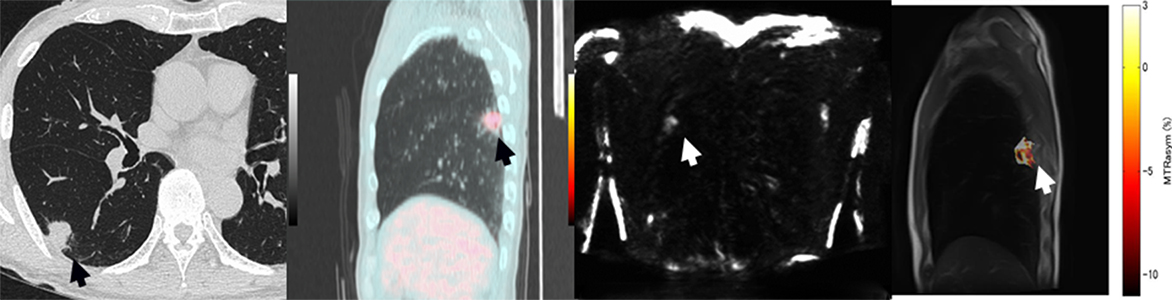
Figure 1. 69-year-old female with invasive adenocarcinoma (L to R: thin-section CT, SUVmax map fused with CT, DWI obtained by FASE sequence and MTRasym map fused with T2WI)
Thin-section CT demonstrates a nodule (arrow) with pleural indentation, notch and spicula in the right lower lobe. This nodule (arows) shows SUVmax as 3.5, ADC as 1.18×10⁻³mm2/s and MTRasym as 1.99%. This case was true-positive on PET/CT, DWI and CEST imaging.
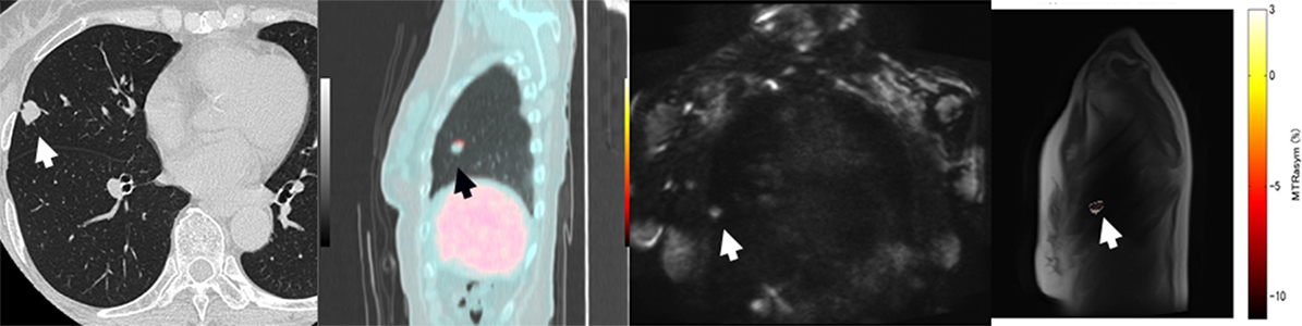
Figure 2. 69-year-old female with organizing pneumonia (L to R: thin-section CT, SUVmax map fused with CT, DWI obtained by FASE sequence and MTRasym map fused with T2WI)
Thin-section CT demonstrates a nodule (arrow) with pleural indentation and notch in the right middle lobe. This nodule (arows) shows SUVmax as 2.5, ADC as 1.28×10⁻³mm2/s and MTRasym as -10.37%. This case was false-positive on PET/CT and true-negative on DWI and CEST imaging.
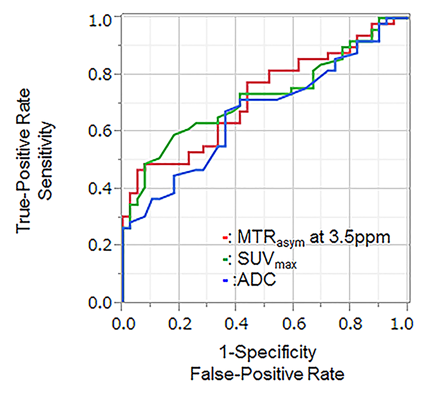
Figure 3. Results of ROC analysis on MTRasym at 3.5ppm, ADC and SUVmax for distinguishing malignant and benign groups. .
There was no significant difference of area under the curve (Az) among MTRasym at 3.5ppm (Az=0.72), ADC (Az=0.67) and SUVmax (Az=0.72) (p>0.05).