4892
Aortic Wall MR Imaging for Assessing Rosuvastatin Therapy in Atherosclerotic Animal Model1Beijing Hospital, Beijing, People's Republic of China
Synopsis
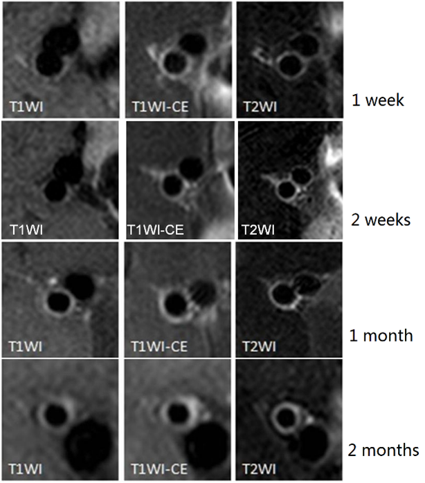
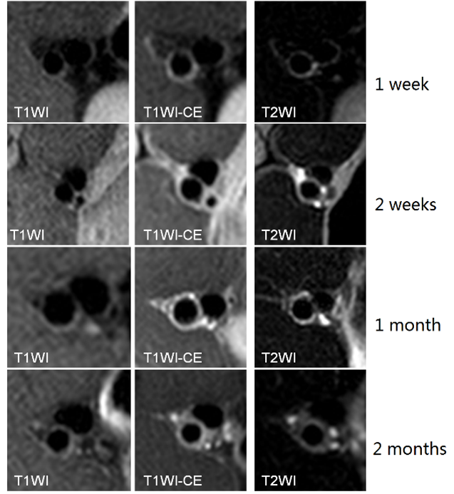
Here we present the high-resolution MRI imaging for assessing therapy of titrated rosuvastatin for atherosclerosis in rabbit models. This study shows that abdominal aorta wall in both control and treated rabbits get thicker with prolonged continuous feeding of cholesterol, aortic plaques grows bigger with this progression. The MRI vascular wall imaging measured atherosclerotic plaques with well correlation of histological classifications. Statical regression showed that the group with rosuvastatin treatment reduced the aortic plaques comparing to the control rabbits.
Introduction
Atherosclerotic plaques pose risks of disruption and thrombosis which leads to most acute cardiovascular events1, 2, it was found that intensive lipid lowering therapy with titrated rosuvastatin yields greater atherosclerotic aortic plaque regression3, 4. Animal models are suitable for comprehensive understanding of this pathology5 and confirm the clinical feasibility of rosuvastatin therapy for atherosclerosis. A modified rabbit model of atherosclerosis was used for MRI exams and detailed histology of rabbit aortic plaques.Methods
80 adult (3 months old, 2.8 kg), male, New Zealand white rabbits were purchased from Animal Models Center. All rabbits were fed a 1% cholesterol diet5 a week prior to balloon injury of the abdominal aorta, half of rabbits will serve as control, and the other half will be treated with rosuvastatin4. 10 rabbits each time were randomly picked 1 week, 2 weeks, 1 month and 2 months after the injury. MRI exams were performed for in vivo aorta exams, followed by aorta extraction. The sections from extraction were then stained with hematoxylin and eosin (HE). All in vivo aorta MRI scans were performed in Philips Intera Achieva 3.0T, with knee coil for higher SNR with small FOV. Each exam included 3D TOF, T1WI, T2WI and dynamic enhanced T1WI sequences. Regression analysis was applied for comparisons between MR aorta wall imaging and the histological studies.Results
48 rabbits completed both MRI and histology exams, with 7 at 1 week, 3 at 2 weeks, 5 at 1 month and 8 at 2 months from control groups, as well as 5, 6, 7 and 6 at each corresponding time from the rabbits treated with rosuvastatin. There was a strong linear correlation between vascular wall area(WA) , wall thickness (WT), lumen area (LA) and total vascular area (TVA) by measured by MRI and histology extractions (r=0.623,P =0.003;r=0.631,P=0.004;r=0.624,P =0.002;r=0.578,P =0.003). Both control and those rabbits treated with rosuvastatin were fed with cholesterol diet. However, the later ones showed reduction in atherosclerotic plaque burden at each exam time, from MRI and histology exams.Discussion and conclusion
High resolution aortic wall imaging at 3 Tesla MRI differentiated and accessed rabbit plaques before and after rosuvastatin treatment. By the measurements of MRI and histology, we firmed that rosuvastatin therapy result in reduction of plaque progression among rabbits with atherosclerosis.Acknowledgements
No acknowledgement found.References
1. Saam T, et al. Predictors of Carotid Atherosclerotic Plaque Progression as Measured by Noninvasive Magnetic Resonance Imaging[J]. Atherosclerosis, 2007,194(2): e34-42.
2. Hayashi K, et al. Variations in atherosclerosis and remodeling patterns in aorta and carotids[J]. J Cardiov Magn Reson, 2010, 12(5):10.
3. Ayaori M, et al. Effects of bezafibrate therapy on atherosclerotic plaques detected by MRI in dyslipidemic patients with hypertriglyceridemia[J]. Atherosclerosis, 2008, 196 (1):425-433.
4. Yogo M, et al. Intensive lipid lowering therapy with titrated rosuvastatin yields greater atherosclerotic aortic plaque regression: Serial magnetic resonance imaging observations from RAPID study. Atherosclerosis.2014 Jan; 232(1):31-9.
5. Phinikaridou A, et al. A robust rabbit model of human atherosclerosis and atherothrombosis[J]. J Lipid Res, 2009. 50(5):787-797.
Figures