4863
Characterization of renal parenchyma impairment in partial unilateral ureteral obstruction in mice with Intravoxel Incoherent Motion MR imaging1INSERM UMR 1149, Paris, France, 2Univ Lyon, INSA‐Lyon, Université Claude Bernard Lyon 1, 3Hôpital Robert Debré, APHP, Université Paris Diderot, Sorbonne Paris Cité, 4Pediatric Surgery and Urology, Hôpital Robert Debré, APHP, Université Paris Diderot, Sorbonne Paris Cité, 5Université Reims Champagne Ardennes,American Memorial Hospital
Synopsis
Ureteropelvic junction obstruction constitutes a major cause of progressive pediatric renal disease. To date the follow-up of patients is difficult because there is a lack of non-invasive biomarkers. Here we propose to quantitatively characterize impairment of the kidney parenchyma after partial unilateral ureteral obstruction (pUUO) on mice using an intravoxel incoherent motion diffusion sequence. The results suggest that an f reduction is associated with a decrease in the volume of the renal parenchyma, which could be related to decreased renal vascularization. The later may occur before impairment by fibrosis and the findings are in accordance with the literature on pUUO.
Introduction
Obstructive nephropathy constitutes a major cause of pediatric renal progressive disease. To date the follow-up of patients is difficult as the progression of the disease is poorly understood (1) and there is a lack of non-invasive biomarkers (2-4). The indication for surgery generally depends on imaging criteria requiring a few months follow-up of patients. The biological mechanism underlying the renal response to obstruction can be investigated using a model of partial unilateral ureteral obstruction (pUUO) in mice. Renal function and kidney morphology data can be evaluated using renal ultrasound, and scintigraphy, and uro-MRI but these methods are poorly linked to histological change and not all are quantitative. We have proven the importance of morphological impairment evaluation by MRI in pUUO. To go further we propose to use intravoxel incoherent motion (IVIM) diffusion sequence to characterize kidney parenchyma impairment.Material and method
The diffusion
coefficient (Dslow), the perfusion coefficient (Dfast) and the perfusion
fraction (f) were extracted from IVIM data acquired on a 7T preclinical system
using a Matlab homemade software. The imaging method was validated on 10 sham
wild type (WT) mice. Then 10 WT mice were subjected to UUOp at day 3 of life.
At day 75, mice underwent MRI examinations with a morphological T2 image and an
IVIM sequence.Results
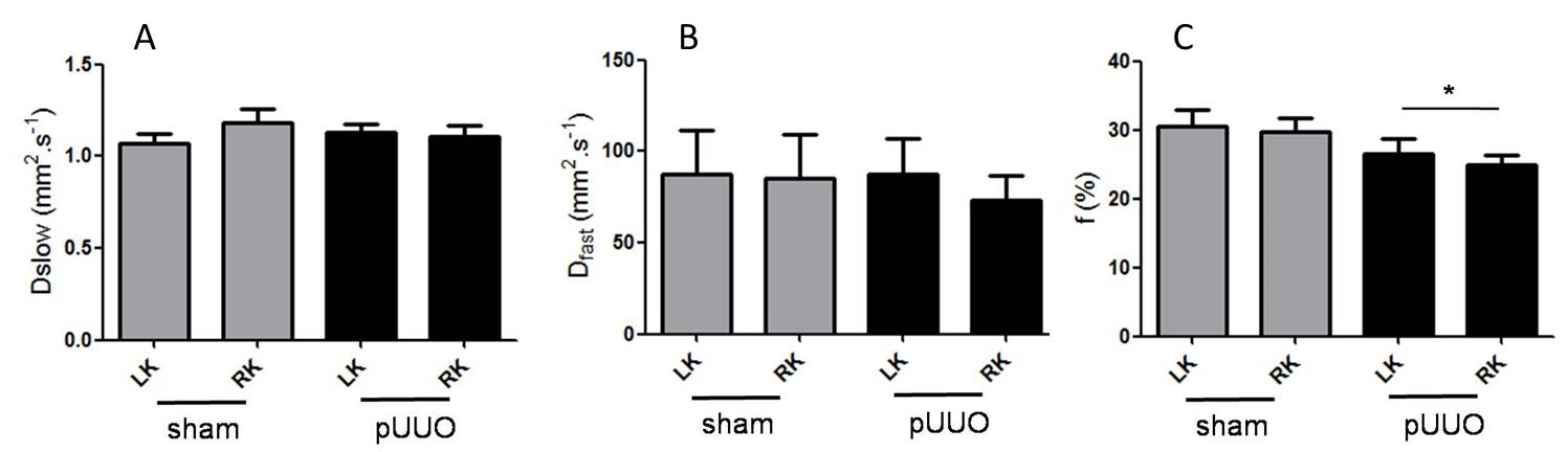
As shown on figure 1, diffusion parameters extracted from IVIM imaging were similar in both kidneys of sham WT mice. Mean values of Dslow, Dfast and f were respectively 1.17±0.22 mm2.s-1, 84.9±73.8 mm2.s-1and 29.7±6.19 % in the right kidney and 1.07±0.16mm2.s-1, 87.6±71.7 mm2.s-1, and 30.6±7.0 % in the left kidney. For pUUO mice a significant decrease of f (24.9±4.7 %) in the right operated kidney compared to the sham right kidney was measured (p=0.04). In 60 % of pUUO mice, characterized by a significant difference between the right and the left kidney, strong correlation between f and the volume of the right kidney was observed (spearman coefficient=0.94, p=0.01).Conclusion
The IVIM sequence has been validated for the first time on mouse kidneys at 7T. According to the literature on UUO mice model and more specifically on pUUO, our study suggests that a f reduction associated with a decrease volume parenchyma could be related to a decrease of renal vascularization, appearing before fibrosis impairment (5-8). Perfusion fraction is a good candidate as a MRI biomarker to follow quantitatively the early changes of kidney pathophysiology in pUUO. These findings are of specific interest in clinical application. All the current investigations detect the impact of obstruction only at the irreversible stage of lost of function secondary to obstruction. In the work of Ichikawa S et al. (9) IVIM examination has already been applied on 365 patients with renal dysfunction and has shown that perfusion fraction could be an earlier and more sensitive marker than molecular diffusion to parenchymal changes. As a perspective we believe that this work may need to be completed by analysis of microperfusion after relieve of obstruction before to be aplied in a multicenter clinical trial.Acknowledgements
The authors thank the American Memorial Hospital Foundation Inc. (Boston USA) for granting a part of this project.References
1. Chevalier RL, Forbes MS, Thornhill BA. Ureteral obstruction as a model of renal interstitial fibrosis and obstructive nephropathy. Kidney international. 2009;75(11):1145-52.
2. Nikken JJ, Krestin GP. MRI of the kidney-state of the art. Eur Radiol. 2007 Nov;17(11):2780-93.
3. Takahashi T, Wang F, Quarles CC. Current MRI techniques for the assessment of renal disease. Curr Opin Nephrol Hypertens. 2015 May;24(3):217-23.
4. Grenier N, Merville P, Combe C. Radiologic imaging of the renal parenchyma structure and function. Nat Rev Nephrol. 2016 Jun;12(6):348-59.
5- Hennedige T, Koh TS, Hartono S, Yan YY, Song IC, Zheng L, et al. Intravoxel incoherent imaging of renal fibrosis induced in a murine model of unilateral ureteral obstruction. Magn Reson Imaging. 2015 Dec;33(10):1324-8.
6- Cheung CM, Shurrab AE, Buckley DL, Hegarty J, Middleton RJ, Mamtora H, et al. MR-derived renal morphology and renal function in patients with atherosclerotic renovascular disease. Kidney Int. 2006 Feb;69(4):715-22.
7- Chevalier RL. Growth factors and apoptosis in neonatal ureteral obstruction. J Am Soc Nephrol. 1996 Aug;7(8):1098-105.8- Chevalier RL. Pathophysiology of obstructive nephropathy in the newborn. Semin Nephrol. 1998 Nov;18(6):585-93.
9- Ichikawa S, Motosugi U, Ichikawa T, Sano K, Morisaka H, Araki T.
Intravoxel incoherent motion imaging of the kidney: alterations in diffusion
and perfusion in patients with renal dysfunction. Magn Reson Imaging. 2013
Apr;31(3):414-7.
Figures