4841
Magnetic Resonance Imaging Features of Immunoglobulin G4 Related Kidney Disease1Radiology, The Fisrt Affiliated Hospital, College of Medicine, Zhejiang University, Hangzhou, People's Republic of China
Synopsis
Immunoglobulin G4 related disease (IgG4-RD) is a recently recognized distinct disease entity that can affect many organs/tissues. The kidney is a frequently involved organ with tubulointerstitial nephritis (TIN), and the renal lesions are collectively referred to as IgG4-related kidney disease (IgG4-RKD). Although definitive diagnosis requires histopathologic analysis, imaging plays a crucial role in demonstrating the involved organs/tissues. We retrospectively reviewed the magnetic resonance (MR) imaging findings of 13 patients diagnosed as IgG4-RKD. Various features were assessed, including renal size, margin between cortex and medulla, signal intensity on T1, T2 and diffusion weighted MR images, contrast enhancement, collecting system and/or renal fascia changes. MR imaging features, such as morphology changes, signal intensity abnormality especially on DWI, collecting system and/or renal fascia involvement enables the diagnosis of IgG4-RKD.
Purpose
To retrospectively investigate the features of immunoglobulin G4 related kidney disease (IgG4-RKD) in magnetic resonance (MR) imaging.
Method
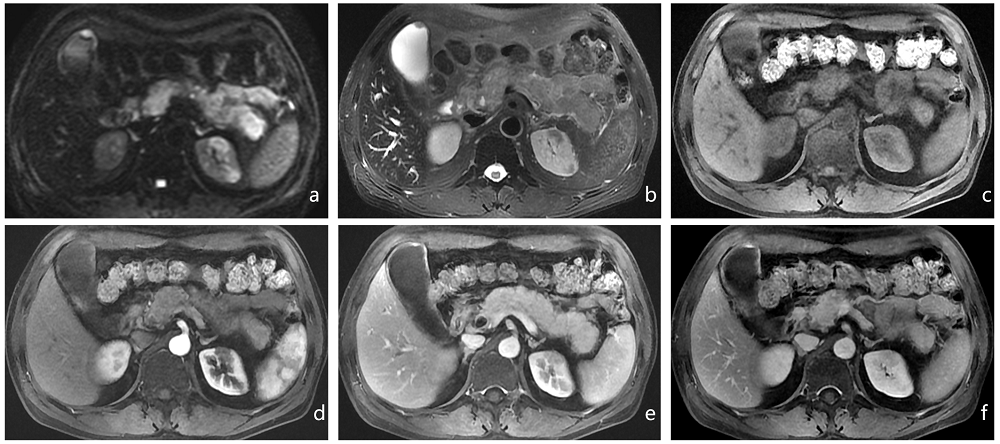
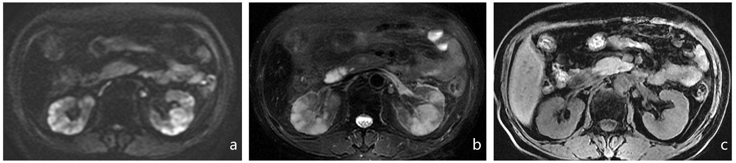
This retrospective study was approved by the institutional review board, and the requirement for informed patient consent was waived. 13 patients with diagnosis of IgG4-RKD were included in the study. All patients underwent MR imaging. Various features were assessed, including renal size, margin between cortex and medulla, signal intensity on T1, T2 and diffusion weighted MR images, contrast enhancement, collecting system and/or renal fascia changes.Results
The kidneys showed diffuse bilateral enlargement (2 patients), bilateral atrophy (2 patients) and segmental atrophy (2 patients). The margin between cortex and medulla were obscure in 12 patients on T2-weighted images. Masslike or patchy lesions were observed in 11 patients, with distribution that could be focal (4 patients), multifocal (7 patients) and involve unilateral kidney (4 patients) or bilateral kidneys (7 patients). All the lesions appeared hyperintense in diffusion weighted imaging (DWI), with hypointense (3 patients) or isointense (8 patients) on T1- weighted imaging ,and hyperintense (2 patients), isointense (7 patients) or hypointense (2 patients) on T2-weighted imaging. After contrast enhancement, all the masslike or patchy lesions appeared mild or moderate progressive enhancement during the corticomedullary, nephrographic and excretory phases. Collecting system involvement was observed in 4 patients, and renal fascia involvement was observed in all patients.Conclusion
MR imaging features like morphology changes, signal intensity abnormality especially on DWI, collecting system and/or renal fascia involvement enables the diagnosis of IgG4-related kidney disease.Acknowledgements
This study was supported by grants from the Zhejiang Provincial Natural Science Foundation of China (LZ16H180001), and partly from the Zhejiang Provincial Natural Science Foundation of China (LY17H160010). The funders had no role in study design, data collection and analysis, decision to publish, or preparation of the manuscript.References
1.Takahashi N, Kawashima A, Fletcher JG, et al. Renal involvement in patients with autoimmune pancreatitis: CT and MR imaging findings. Radiology. 2007;242(3):791-801.
2.Kim B, Kim JH, Byun JH, et al. IgG4-related kidney disease: MRI findings with emphasis on the usefulness of diffusion-weighted imaging. Eur J Radiol. 2014;83(7):1057-62.
3.Dauvergne M, Moktefi A, Rabant M, et al. Membranous Nephropathy Associated With Immunological Disorder-Related Liver Disease: A Retrospective Study of 10 Cases. Medicine. 2015;94(30):e1243.
Figures