4839
The role of Multiparametric prostate MRI in the detection, biopsy, and staging of prostate cancer.1Radiology, Northwell Health, Manhasset, NY, United States
Synopsis
Prostate cancer is the second cause of cancer related death in men. Historically, screening has consisted of serum PSA, digital rectal exams, and random US guided transrectal biopsy. Compared to older methods, mp-MRI is superior for the detection and staging of prostate cancer due to its improved visualization and lesion characterization. The PI-RADS classification system is a schema developed concordantly with mp-MRI in order to better characterize the clinical significance of imaging findings. Increasingly, mp-MRI is taking a central role in detection, staging and biopsy of prostate neoplasm. In the future it may emerge as a primary screening tool.
Purpose:
To describe the evolving role of prostate MRI in the detection, staging, and biopsy of prostate cancer through discussion of prostate MRI imaging techniques, review of anatomy and imaging characteristics of malignancy, and highlighting fusion biopsy techniques.Outline of Content:
1. Prostate cancer epidemiology, review of traditional screening and detection techniques
2. Discussion of mp-MRI of the prostate as an evolving method for detection and local staging
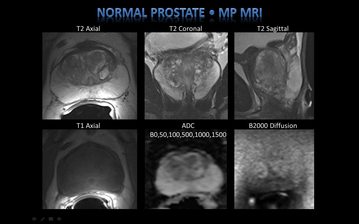
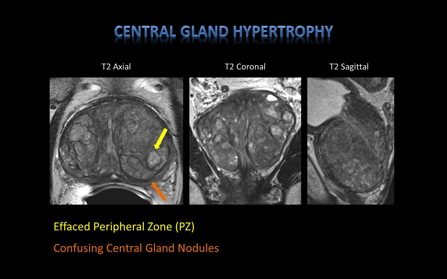
3. Review of prostate gland anatomy and typical imaging appearance on MRI
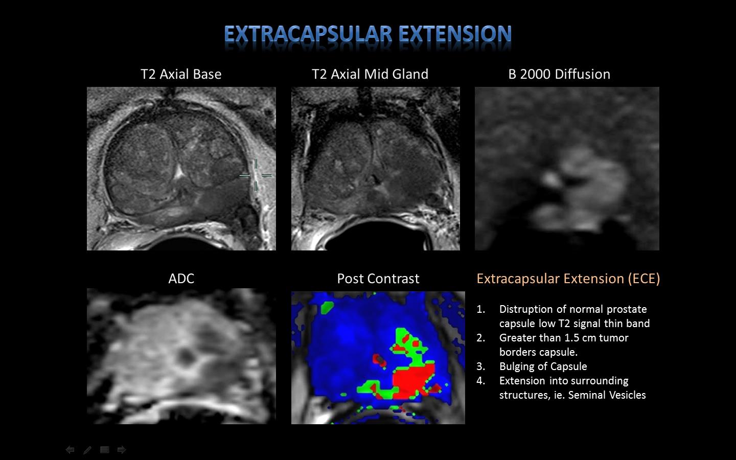
4. Depiction of imaging characteristics of malignancy
5. Description and overview of lesion reporting with PI-RADS classification
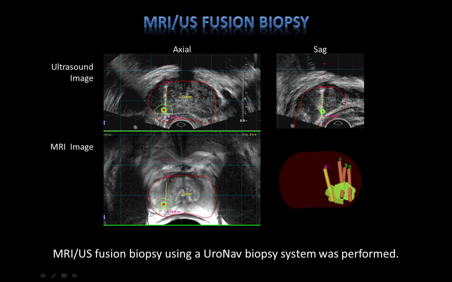
6. Overview of role of MRI in US fusion biopsy to obtain localized and accurate sampling of identified lesions.
7. Future roles of mp-MRI in prostate cancer screening
8. Summary
Acknowledgements
1. Eran Ben-Levi, MD, System Chief of Body Imaging, Long Island Jewish Medical Center.
2. Ardeshir R Rastinehad, DO, Associate Professor Urology, Associate Professor Radiology, The Mount Sinai Hospital.
References
1. Hoeks CMA, Barentsz JO, Hambrock T, Yakar D, Somford DM, Heijmink SWTPJ, et al. Prostate cancer: multiparametric MR imaging for detection, localization, and staging. Radiology 2011;261(1):46–66.D.
2. Bonekamp, M. A. Jacobs, R. El-Khouli, D. Stoianovici, K. J. Macura, Advancements in MR imaging of the prostate: From diagnosis to interventions. Radiographics 31, 677–703 (2011).
3. Costa DN, Pedrosa I, Donato F Jr et al: MR imaging-transrectal US fusion for targeted prostate biopsies: implications for diagnosis and clinical management. Radiographics 2015; 35: 696.
4. Yacoub JH, Verma S, Moulton JS, Eggener S, Aytekin O. Imaging-guided prostate biopsy: conventional and emerging techniques. Radiographics 2012; 32: 819–837
5. Marks L, Young S, Natarajan S. MRI-ultrasound fusion for guidance of targeted prostate biopsy. Curr Opin Urol 2013;23:43–50.
6. Rosenkrantz AB, Verma S et al. Prostate Magnetic Resonance Imaging and Magnetic Resonance Imaging Targeted Biopsy in Patients with a Prior Negative Biopsy: A Consensus Statement by AUA and SAR. J Urol. 2016 Jun 16.
7. Rosenkrantz, AB, Babb JS et al. Proposed Adjustment to PI-RADS Version 2 Decision Rules: Impact on Prostate Cancer Detection. Radiology, 2016 Oct 26:161124.
8. Toner L, Papa N et al. Multiparametric magnetic resonance imaging for prostate cancer-a comparative study including radical prostatectomy specimens.World J Urol. 2016 Oct 26.
9. Kim W, Kim CK et al. Evaluation of extracapsular extension in prostate cancer using qualitative and quantitative multiparametric MRI. J Magn Reson Imaging. 2016 Oct 17.
10. Barrett T, Haider MA. The Emerging Role of MRI in Prostate Cancer Active Surveillance and Ongoing Challenges. AJR Am J Roentgenol. 2016 Oct 11:1-9.
11. Rosenkrantz AB, Taneja SS. Radiologist, be aware: ten pitfalls that confound the interpretation of multiparametric prostate MRI. AJR Am J Roentgenol 2014;202:109e20.
12. Barrett T, Turkbey B, Choyke PL. PI-RADS version 2: what you need to know. Clin Radiol 2015;70:1165e76.
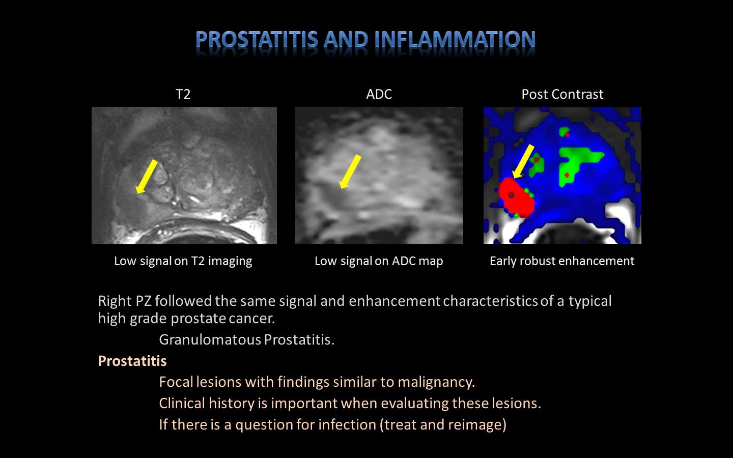
13. Yu J, Fulcher AS, Turner MA, Cockrell CH, Cote EP, Wallace TJ. Prostate cancer and its mimics at multiparametric prostate MRI. Br J Radiol 2014; 87:20130659.
Figures