4831
Compressed Sensing Black Blood SPACE for Abdominal Aortic Aneurysmal Vessel Wall Imaging1Siemens Healthcare, San Francisco, CA, United States, 2Radiology and Biomedical Imaging, UCSF, San Francisco, CA, United States, 3Siemens Healthcare, Erlangen, Germany, 4UCSF, San Francisco, CA, United States
Synopsis
Abdominal aortic vessel wall imaging has been interested and used for studying pathological vasculature. In this study, compressed sensing (CS) 3D SPACE DANTE technique is proposed for imaging abdominal aortic aneurysm (AAA) vessel wall to accelerate imaging acquisition. Its scan/reconstruction parameters were optimized on normal volunteers then, used for patient scan. DANTE provided sufficiently dark blood signal at a large vessel like the aorta, which is essential to delineate the vessel wall. CS SPACE DANTE provided comparable image quality and vessel wall-lumen signal contrast as compared to non-accelerated SPACE DANTE technique.
Introduction
MR abdominal aortic vessel wall imaging has gained interest for studying vascular pathology such as thrombus and aneurysm (1). 3D SPACE DANTE (Delayed Alternating with Nutation for Tailored Excitation) (2) has been proposed to provide high-resolution 3D images of abdominal vasculature with dark blood signal for characterizing aneurysmal wall and intra-luminal thrombus (3). However, it still requires a relatively long scan time (~ 7min) to produce quality images with good aortic luminal contrast. Although it is clinically feasible, shortened acquisition would always be of benefit to image quality (by less chance of motion contamination), patient comfort, and imaging throughput. In this paper, we propose the SPACE DANTE with compressed sensing (CS) technique to perform abdominal aortic aneurysm (AAA) vessel wall imaging in more clinically feasible scan time. Imaging was accelerated by sparse and incoherent under-sampling and a nonlinear iterative reconstruction scheme was used for image formation (4,5,6).Methods and Materials
MR acquisition was performed on a 3T system (MAGNETOM Skyra, Siemens, Erlangen, Germany) using a spine array coil and anterior body array coil. Two normal subjects and one patient with AAA were scanned using T1-weighted 3D SPACE DANTE (prototype) and 3D CS SPACE DANTE (prototype). CS scan and its online reconstruction parameters were optimized on the normal subjects and then, used for the patient scan. The coronal imaging slab was placed to cover the abdominal aorta from a renal artery to an aorta bifurcation. The acquisition parameters are as follows: TE/TR=20/814 msec, 1.3 mm isotropic resolution, 6/8 slice partial Fourier (only for non-CS acquisition), 67.6 mm slice coverage in coronal orientation with 15.4% slice oversampling, fat saturation pulse, 60 echo train length, no respiratory or cardiac gating. For comparison purpose, three protocols were run on the normal volunteers; (1) SPACE DANTE for 7:10 min scan (2) SPACE DANTE with GRAPPA 3 acceleration for 2:48 min scan, and (3) CS SPACE DANTE (20% under-sampling, 20 iterations, 0.002~0.004 regularization factor) for 2:48 min scan. Following parameters were used for DANTE preparation: FA=150, 100-pulse train, 1-msec inter-pulse duration, and 15,000 mT/m*usec spoiling gradient pulse along all three axes.Results
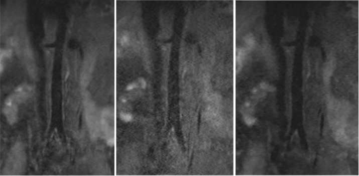
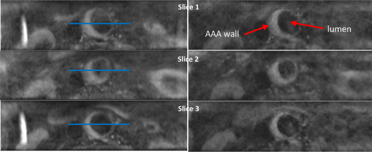
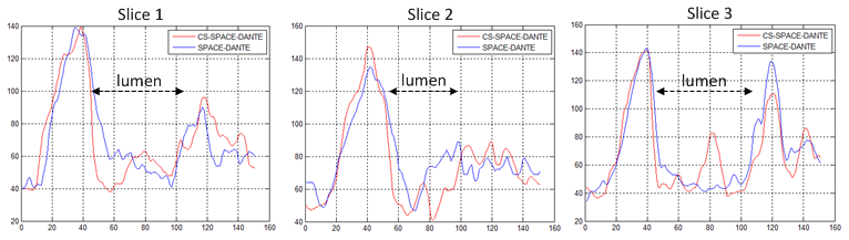
Figure 1 shows acquired coronal images at the aorta bifurcation from a normal subject. The image quality of CS SPACE DANTE with parameter optimization (right) is shown to be comparable to that of SPACE DANTE (left) with 2.6 times faster acquisition, while SPACE DANTE with GRAPPA 3 (center) showed poorer image quality. Figure 2 shows axial reformat of AAA image of the patient (left: SPACE DANTE and right: CS SPACE DANTE). CS SPACE DANTE clearly depicts the AAA and its inner and outer wall boundaries well enough for the further segmentation. Vessel wall-lumen signal contrast of both scans was comparable in that the wall signal is clearly distinguishable from the lumen signal (Figure 3). Overall, DANTE provided dark blood signal at a large vessel like the aorta in both scans, which is essential to delineate the vessel wall. Increased signal in the vessel lumen in the slice 3 from CS SPACE DANTE in Figure 3 is caused by regionally less suppressed blood signal, which may occur depending on the blood flow condition in a large vessel.Discussion and Conclusion
Accelerated 3D black blood AAA imaging using compressed sensing technique was proposed to reduce acquisition time, yet largely maintaining image quality. This may help reduce motion artifacts, increase patient comfort, and increase scan throughput. Although highly under-sampled acquisition can lead to artifacts such as blurring or contrast washout, the CS scan/reconstruction parameters can be optimized in order to provide application-specific image quality. In this work, CS SPACE DANTE was shown to provide comparable image quality and vessel wall-lumen signal contrast to the conventional SPACE DANTE with much shortened acquisition time. Although more clinical validation is needed, CS SPACE DANTE shows potential to be a viable tool for 3D black blood AAA wall imaging.Acknowledgements
The authors would like to acknowledge Jens Wetzl and Michael Zenge for their work on the image reconstruction framework.References
(1) Nguyen et al, Eur J Vasc Endovasc Srg 2014;48:676-684 (2) Li et al, MRM 2012;68:1423-1438 (3) Zhu et al, MRI 2016;34:18-25 (4) Stalder et al, MRM 2015;74(6):1652-60 (5) Liang et al, MRM 2009;62:1574-1584 (6) Fritz et al, Inv Rad 2016;51(6):400-9
Figures