4819
Radial-SWI in Human Fetal Imaging1Department of Biomedical Engineering, Wayne State University, Detroit, MI, United States, 2Department of Radiology, Wayne State University, Detroit, MI, United States, 3Department of Obstetrics and Gynecology, Wayne State University, Detroit, MI, United States, 4Perinatology Research Branch, NICHD/NIH/DHHS, Bethesda, MD, United States
Synopsis
Radial-SWI in human fetal imaging is presented which includes: (a) determining the minimum number of radial projections necessary for fast measurement of intravascular phase in the blood vessels in human adult and fetus, without loss of accuracy; and (b) exploring the feasibility of fetal venography using radial-SWI. Results of this study illustrate that (a) in both fetal and adult imaging, accurate quantification of intravascular phase from the superior-sagittal-sinus is possible from radial SWI with just 161 projections, and (b) venograms in fetal brain were presented using radial-SWI.
Introduction
Susceptibility-weighted-imaging (SWI), a fully flow compensated gradient echo sequence (GRE), has been used previously to: a) quantify blood oxygenation1 from intravascular phase; and b) to generate venograms2 in the human fetus. However, its consistent application is a challenge due to fetal motion. Radial trajectories, on the other hand, sample the centre of k-space densely and hence are inherently insensitive to motion3. However, it takes approximately 1.5 times longer to collect data radially compared to a Cartesian trajectory for the same resolution4. In this work, our aims were: (a) to determine the minimum number of radial projections necessary for fast measurement of intravascular phase in the blood vessels in human adults and fetuses, without loss of accuracy; and (b) to present the feasibility of fetal venography using radial-SWI.Material and Methods
All the MRI scans were performed at 3.0T (Siemens, Verio, Erlangen, Germany). Aim-1: Assessing accuracy of intravascular phase as a function of the number of radial projections (Np): A straw, filled with gadolinium (1.45mM) was placed in a cylindrical container filled with distilled water and was scanned using following parameters: fully flow compensated 2D GRE (radial-SWI) with sequential radial spoke sampling, readout points (Nr)=720, Np=641, TE=15ms, TR=288ms, flip angle (FA)=27°, resolution=0.35×0.35×3mm3, bandwidth (BW)=100 Hz/pixel. Cartesian SWI data was also acquired with the same timing parameters as a control dataset to ascertain accuracy of phase reconstruction from radial-SWI. After obtaining the informed consent in accordance with the local institutional review board guidelines, data were also acquired from a healthy adult volunteer and an adult pregnant subject in the third trimester with normal, singleton pregnancy. The radial-SWI parameters used in the adult head were: Nr=720, Np=641 (over 360°), TE =15 ms, TR=100ms, FA = 30°, resolution=0.4×0.4×3mm3, BW=101Hz/pixel. Radial-SWI in the human fetus (GA:29 weeks) had the following parameters: Nr=720, Np =361, TE=15ms, TR=100ms, FA=200, resolution=0.5×0.5×3mm3, BW=120Hz/pixel. Data was undersampled azimuthally by a factor of 2, 4 and 8 and the phase image was filtered using a 48x48 homodyne filter. Relative phase with respect to the background region (∆φ) was measured within the straw or the superior sagittal sinus (SSS) vein, in the case of adult and fetal brain. The region-of-interest (ROI) contained at least 15 voxels. Aim-2: Feasibility of fetal venography: The fetal radial-SWI data was used for generating venograms from 2 slices and 4 phase multiplications with a sliding window approach.Results
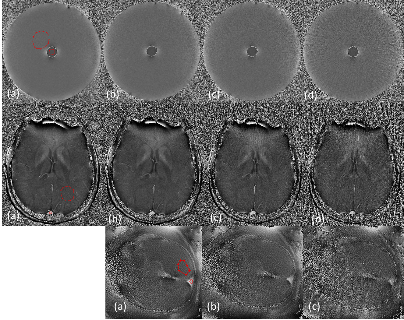
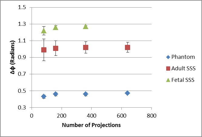
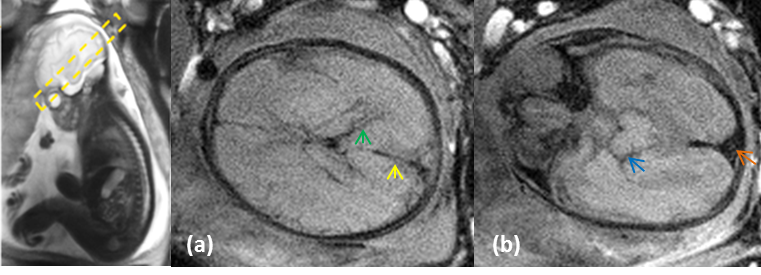
The phase ∆φ obtained from radial and Cartesian images of the phantom were 0.47±0.09 and 0.46±0.08 radians, respectively confirming the consistency of the radial reconstruction. Figure-1 shows phase images of a phantom and the adult brain obtained from 641, 361, 161 and 81 projections and the fetal brain from 361,161 and 81 projections. The measured ∆φ versus the number of spokes is plotted in Figure-2. With a decreasing number of spokes, the measured phase remains unchanged until 161 spokes, with a relative error <2.5%. The corresponding measurement error, however, increases due to increasing under-sampling artifacts. Figure-3 shows the fetal vessels (as shown by arrows) such as the basal vein of Rosenthal (green), middle atrial veins (blue), SSS(orange) and straight-sinus (yellow) in the venograms.Discussion
Since ∆φ obtained in the phantom was the same in the Cartesian and radial acquisitions, we took 641 radial projections as our reference for the radial subsampling experiment. As the undersampling was increased, streaking artifacts got more pronounced. In terms of ∆φ quantification, however, the measurement remains reliable until 161 projections in all the cases studied. The standard error increased significantly and ∆φ began showing bias error at 81 projections. Since, the radial acquisition was in sequential order, exploring fractional undersampling factors was not possible. The Golden Angle5 sampling scheme could offer further freedom in this regard and provide a finer determination of the undersampling threshold for accurate intravascular phase determination. Furthermore, we present the first radial-SWI application on the human fetus. Because parallel imaging was not taken advantage of in this work, the current radial-SWI technique takes longer compared to Cartesian SWI, depending on the number of spokes acquired. With parallel imaging and the use of approaches like radial-EPI6 sampling with flow compensation for each readout, radial-SWI scan time can be further reduced considerably.Conclusion
This is the first study that shows the feasibility of generating venograms in the human fetal brain using radial-SWI. In both fetal and adult imaging, accurate quantification of intravascular phase from the SSS vessel is possible from radial SWI with a minimum of 161 projections.Acknowledgements
No acknowledgement found.References
1. Neelavalli, Jaladhar, et al. Measuring venous blood oxygenation in fetal brain using susceptibility-weighted imaging. Journal of Magnetic Resonance Imaging 39.4 (2014): 998-1006.
2. Neelavalli, Jaladhar, et al. MR venography of the fetal brain using susceptibility weighted imaging. Journal of Magnetic Resonance Imaging 40.4 (2014): 949-957.
3. Block, Kai Tobias, Martin Uecker, and Jens Frahm. Undersampled radial MRI with multiple coils. Iterative image reconstruction using a total variation constraint. Magnetic resonance in medicine 57.6 (2007): 1086-1098.
4. Haacke, E. Mark, et al. Magnetic resonance imaging: physical principles and sequence design. Vol. 82. New York:: Wiley-Liss, 1999.
5. Winkelmann, Stefanie, et al. An optimal radial profile order based on the Golden Ratio for time-resolved MRI. IEEE transactions on medical imaging 26.1 (2007): 68-76.
6. Winkelmann, Stefanie, et al. Simultaneous imaging and R2* mapping using a radial multi-gradient-echo (rMGE) sequence. Journal of Magnetic Resonance Imaging 24.4 (2006): 939-944.
Figures