4816
MR non-Gaussian diffusion model of female cervix: a diffusion kurtosis imaging studyWenhui Guo1, Kuang Fu1, and Lizhi Xie2
1Department of MR, The Second Affiliated Hospital of Harbin Medical University, Harbin, People's Republic of China, 2GE Healthcare, MR Research China, BeiJing, People's Republic of China
Synopsis
To assess the fitted parameters of DKI in female cervix and to investigate their potential in distinguishing tumors from diverse healthy tissues. The presence of rich collagen and fibers in healthy cervix helps to differentiate itself from the heterogeneious and cell-rich malignancies. It is concluded that DKI can be performed as a feasible technique to depict the real phenomenon of non-Gaussian water diffusion behavior in female cervix.
Purpose
DKI provides an estimate for the excess kurtosis of the diffusion displacement probability distribution and is able to characterize the non-Gaussian behavior of water diffusion in vivo. Therefore, the current study aimed to investigate and compare DKI parameters in cervical normal tissues to those of the tumorous tissues.Material and Methods
Thirty females were enrolled in the current study, including 17 healthy volunteers and 13 patients with biopsy-proven well or moderately differentiated uterine cervical squamous cell cancers. All the subjects underwent MR examinations on a 3T MR scanner (Discovery MR750W, GE Healthcare, USA). 4 healthy volunteers were excluded due to the vague boundaries of the cervix stroma. A 2D oblique axial plane protocol (3b-values: 0, 1000, 2000s/mm2, 30 diffusion directions, nex=2), perpendicular to cervical mucus line(in healthy), was applied for DKI acquisition. Nex=2 protocol was introduced to shorten the examination time to prevent movements of the subjects during acquisition period. Images analyses were performed on GE AW4.6 Workstation. Irregular regions of interest (ROIs) were manually drawn to obtain quantitative parameters, such as fractional anisotropy(FA), mean diffusivity (MD), axial diffusivity(Da), radial diffusivity(Dr), mean kurtosis(MK), axial kurtosis(Ka), and radial kurtosis(Kr). According to the fat suppression T2 weighted imaging, ROIs were drawn along with the boundaries of the cervical stroma and the smooth muscle tissues in normal tissues, while the ROIs were drawn encompassed the whole area of the lesions in tumorous tissues. Examples of ROI placement and maps were displayed in Figure.1 and Figure.2. 1-sample K-S test, Paired T-test, independent sample T-test and Pearson’s correlation were applied to investigate the statistical significance of the results.Results
Seven DKI parameters of the tumors and the smooth muscle tissues showed significant correlation(P<0.05) between themselves in each group. However, no correlation was observed between FA and other parameters in the cervical stroma. MD, Da, Dr, MK, Ka, Kr derived from DKI were significantly different between cervical stroma and the smooth muscle (P<0.05). All the parameters except for Kr demonstrated significant difference between cervical stroma and carcinoma. Carcinoma demonstrated decreased FA, MD, Da, Dr than those of the healthy, whereas increased MK, Ka, Kr than those of the cervical stroma and muscle, as shown in Figure 3.Discussion
Female cervix is a cylindrical anatomical structure with massive density of fibrous connective tissues, which is composed of central canal, cervical stroma and few smooth muscles, in continuity with the lower uterus and upper vagina. In the peripheral area of the cervix, ligaments appear to insert into the smooth muscles, such as cardinal ligament and uterosacral ligament, where they display higher signal than that of the cervical stroma in T2WI. Diffusion metrics of the muscles were higher than stroma and tumor, while the kurtosis metrics appeared to be the lowest among the groups. A possible explanation is that water mocleculs are more free to diffuse in fiber tissues. In malignancies, the cell density, microcirculation, membranes permeability and tissue heterogeneity differ from the those in the healthy tissues, which will lead to restricted movement of the water molecules.Conclusion
Diffusion metrics and kurtosis metrics derived from DKI distinguished between the tumorous and normal tissues, as well as among different tissues in the healthy subjects. Therefore, DKI could be a feasible technique to depict the real phenomenon of non-Gaussian water diffusion behavior in female cervix, especial for clinical diagnostic purpose.Acknowledgements
No acknowledgement found.References
[1] Cauter S V, et al. Gliomas: Diffusion Kurtosis MR Imaging in Grading[J]. Radiology, 2012, 263(2):492-501.[2] Rosenkrantz A B, et al. Prostate cancer: feasibility and preliminary experience of a diffusional kurtosis model for detection and assessment of aggressiveness of peripheral zone cancer.[J]. Radiology, 2012, 264(1):126-35.
[3] Sun K, Chen X, Chai W, et al. Breast Cancer: Diffusion Kurtosis MR Imaging-Diagnostic Accuracy and Correlation with Clinical-Pathologic Factors.[J]. Radiology, 2015, 277(1):46-55.
Figures
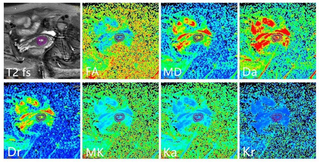
Figure.1Transverse T2-weighted imaging with fat
saturation, FA, MD, Da, Dr, MK, Ka and Kr maps of a 45-year-old healthy woman
were demonstrated. Two kinds of ROIs were drawn along with the cervical stroma
and the smooth muscle tissues.
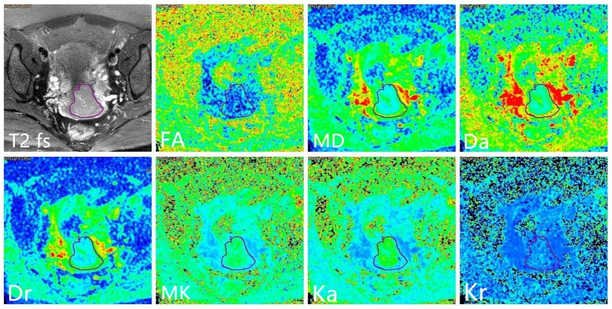
Figure.2 Transverse T2-weighted imaging with fat
saturation, FA, MD, Da, Dr, MK, Ka and Kr maps of a 28-year-old female with cervical
squamous cell cancer. ROI was drawn
encompassed the whole area of the lesion.

Figure.3 Differences of FA, MD, Da, Dr, MK, Ka, Kr among cancer, smooth muscle tissue and cervical stroma.