4814
Red degeneration of uterine leiomyoma: Clinical utility of susceptibility-weighted MR imagingMayumi Takeuchi1, Kenji Matsuzaki2, and Masafumi Harada1
1Department of Radiology, Tokushima University, Tokushima, Japan, 2Department of Radiological Technology, Tokushima Bunri University, Sanuki-city, Japan
Synopsis
Red degeneration of uterine leiomyoma (RDL) is hemorrhagic infarction caused by peripheral venous thrombosis. Peripheral high intensity rim on T1WI due to methemoglobin of blood products confined to thrombosed vessels is characteristic, however, it may not be observed at acute phase. We evaluated MR images including SWI of 17 RDL and 12 usual leiomyomas (UL). High intensity rim on T1WI, low intensity rim on T2WI and on SWI were observed in 47%, 47%, and 100% of RDL, whereas 0%, 8%, and 0% of UL, respectively. SWI may be helpful for the diagnosis of RDL in distinguishing from UL or sarcomas.
Background and purpose of the study
Red degeneration of uterine leiomyoma is a subtype of degeneration that often occurs during pregnancy, or with use of oral contraceptives. On gross pathological examination, it is characterized by a red, hemorrhagic appearance of leiomyoma (Fig. 1). Red degeneration is hemorrhagic infarction caused by venous thrombosis within the periphery of leiomyomas. Characteristic signal intensity patterns have described on MR imaging1, 2 as peripheral high intensity rim on T1-weighted images due to T1-shortening effects of methemoglobin of blood products confined to thrombosed numerous dilated vessels surrounding the tumor (Fig. 1, 2). On T2-weighted images, it may show variable signal intensity with low intensity rim due to T2*-shortening effects of deoxyhemoglobin of blood products confined to thrombosed vessels (Fig. 1, 2). However high intensity rim on T1-weighted images may not be observed at acute phase because of insufficient conversion of deoxyhemoglobin into methemoglobin, low intensity rim on T2-weighted images due to deoxyhemoglobin may be observed even at acute phase (Fig. 3). Because usual leiomyomas show low intensity on T2-weighted images, peripheral rim-like low intensity may also be observed in degenerated leiomyoma without hemorrhagic infarction (Fig. 4). SW imaging can maximize sensitivity to susceptibility effects and has exquisite sensitivity to blood products3-6. We hypothesized that low intensity rim due to deoxyhemoglobin may be more clearly demonstrated on SW imaging and be helpful for the early diagnosis of red degeneration. The purpose of this study was to evaluate the capability of SW imaging for the diagnosis of red degeneration of uterine leiomyomas.Materials and methods
Surgically proven 17 leiomyomas with red degeneration in 11 women who had undergone MRI examinations including SW imaging before surgery were retrospectively evaluated. Fast spin-echo T2-weighted images, gradient-echo T1-weighted images with fat saturation, and SW imaging (SWI: Susceptibility-weighted imaging or SWAN: T2 Star Weighted ANgiography) were obtained for all patients with 3T superconducting MRI system (Discovery MR750, GE), or with 1.5T superconducting MRI system (Signa HDx, GE). Two radiologists with 25 and 16 years of experience in body MRI qualitatively evaluated the images for the presence of high intensity rim on T1-weighted images, and low intensity rim on T2-weighted images and on SW images. The reviewers examined all MR images of the cases independently and then resolved discrepancies by consensus. 12 usual leiomyomas without red degeneration were also evaluated as comparison.Results and discussions
High intensity rim on T1-weighted images was observed in 8 of 17 leiomyomas with red degeneration (47%) and none of 12 usual leiomyomas. The other 9 leiomyomas with red degeneration (53%) showed totally high intensity and rim-like high intensity was not clearly demonstrated. Low intensity rim on T2-weighted images was observed in 8 of 17 leiomyomas with red degeneration (47%), whereas 1 of 12 usual leiomyomas (8%) showed rim-like low intensity. Low intensity rim on SW images was observed in all 17 leiomyomas with red degeneration (100%) and none of 12 usual leiomyomas (Fig. 2-4). Leiomyomas with red degeneration may show diffuse or peripheral high intensity rim on T1-weighted images depending on the degree of intra-tumoral hemorrhage, coagulative necrosis, or hyalinized degeneration1, 2. Peripheral high intensity rim reflecting methemoglobin of blood products confined to thrombosed vessels is characteristic, however, may be observed at subacute phase of red degeneration. High-grade malignant uterine tumor such as leiomyosarcoma may also show diffuse high signal intensity due to massive hemorrhagic necrosis (Fig. 5), so diagnosis of tumors exhibiting diffuse high intensity on T1-weighted images may be problematic. Peripheral low intensity rim on T2-weighted images reflecting venous thrombosis is also characteristic and may be observed even at acute phase due to deoxyhemoglobin, however, usual leiomyoma itself may show low intensity and evaluation of low intensity rim may occasionally be difficult (Fig. 4, 5). In our study the prevalences of high intensity rim on T1-weighted images and low intensity rim on T2-weighted images were 47% and 47%, respectively. SW imaging is a sensitive MRI technique which demonstrates hemorrhage of varying chronicity7, and could reveal peripheral venous thrombosis of leiomyomas with red degeneration as low intensity rim in all cases of the current study.Conclusions
SW imaging is a sensitive MR technique for blood products, and could demonstrate characteristic peripheral low intensity rim of leiomyomas with red degeneration. We conclude that SW imaging may be helpful for the diagnosis of leiomyomas with red degeneration in distinguishing from usual leiomyomas or malignant uterine tumors with massive hemorrhagic necrosis without the use of contrast agent.Acknowledgements
No acknowledgement found.References
1) Kawakami S, Togashi K, Konishi I, et al. Red degeneration of uterine leiomyoma: MR appearance. J Comput Assist Tomogr 1994;18:925-928. 2) Murase E, Siegelman ES, Outwater EK, et al. Uterine leiomyomas: histopathologic features, MR imaging findings, differential diagnosis, and treatment. Radiographics 1999;19:1179-1197. 3) Haacke EM, Xu Y, Cheng YC, et al. Susceptibility weighted imaging (SWI). Magn Reson Med 2004;52:612-618. 4) Boeckh-Behrens T, Lutz J, Lummel N et al. Susceptibility-weighted angiography (SWAN) of cerebral veins and arteries compared to TOF-MRA. Eur J Radiol 2012;81:1238-1245. 5) Sehgal V, Delproposto Z, Haddar D, et al. Susceptibility-weighted imaging to visualize blood products and improve tumor contrast in the study of brain masses. J Magn Reson Imaging 2006;24:41-51. 6) Löbel U, Sedlacik J, Sabin ND, et al. Three-dimensional susceptibility-weighted imaging and two-dimensional T2*-weighted gradient-echo imaging of intratumoral hemorrhages in pediatric diffuse intrinsic pontine glioma. Neuroradiology 2010; 52:1167-1177. 7) Takeuchi M, Matsuzaki K, Harada M. Susceptibility-weighted Imaging of Ovarian Torsion: A Case Report. Magn Reson Med Sci. 2015;14:355-358.Figures
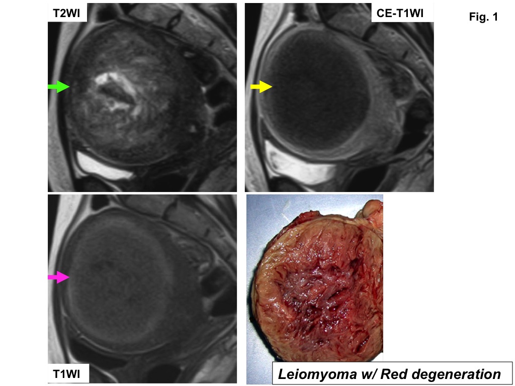
Leiomyoma
with red degeneration (typical). On T1-weighted image, peripheral high
intensity rim (pink arrow) is clearly demonstrated. On T2-weighted image,
peripheral low intensity rim (green arrow) surrounding heterogeneous signal
intensity mass is revealed. On post-contrast T1-weighted image, the mass shows
totally no contrast-enhancement (yellow arrow) due to hemorrhagic infarction of
the tumor. The cut surface of the resected specimen shows red, hemorrhagic
appearance.
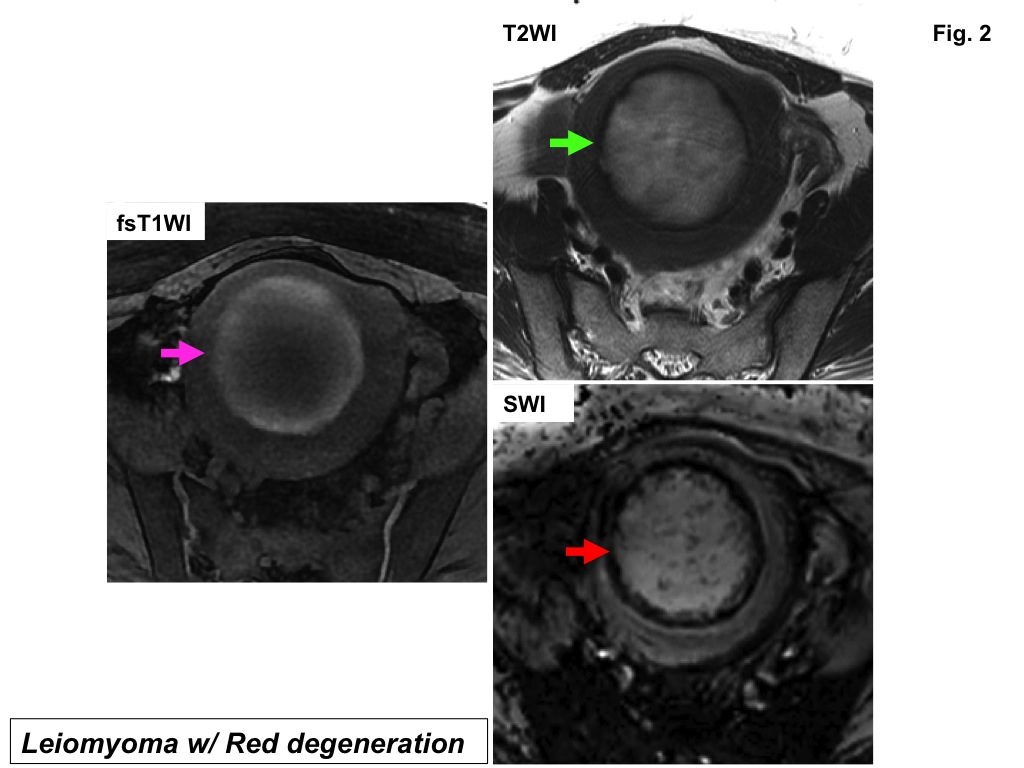
Leiomyoma with red degeneration (typical). On fat-saturated T1-weighted image, peripheral
high intensity rim (pink arrow) is clearly demonstrated. On T2-weighted image,
peripheral low intensity rim (green arrow) surrounding high intensity mass is
revealed. On SW image (SWI), peripheral low intensity rim (red arrow) is
prominent compared with that on T2-weighted image.
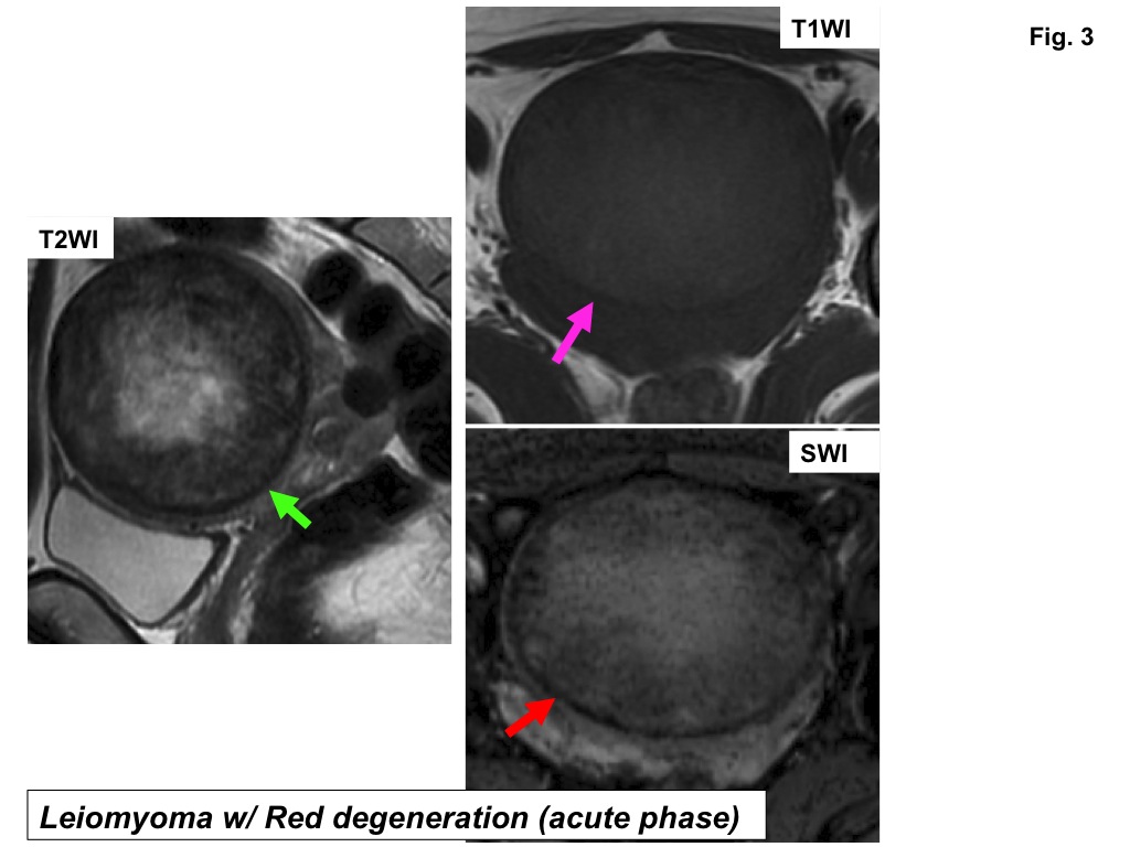
Leiomyoma with red degeneration (acute phase). On T1-weighted image, peripheral high intensity
rim is not demonstrated (pink arrow). On T2-weighted image, peripheral low
intensity rim (green arrow) surrounding heterogeneous signal intensity mass is
revealed. On SW image (SWI), peripheral low intensity rim (red arrow) is clearly
demonstrated.
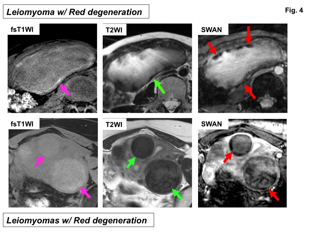
Leiomyomas with red degeneration. In the upper case, large subserosal mass shows discontinuous
high intensity rim on fat-saturated T1-weighted image, and discontinuous low
intensity rims on T2-weighted image and on SW image (SWAN). In the lower case,
intramural masses show totally slight high intensity without definite high
intensity rim on fat-saturated T1-weighted image, and totally low intensity
without definite low intensity rim on T2-weighted image. Discontinuous low
intensity rims are revealed on SW image (SWAN).
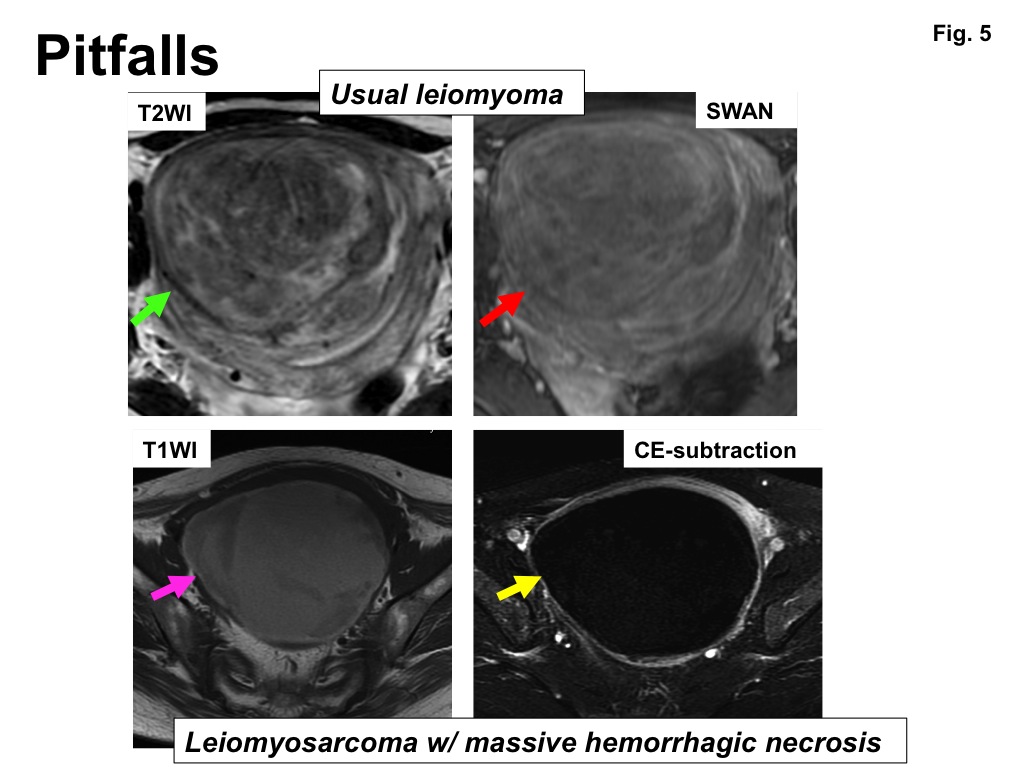
Two pitfall cases. In the upper case (usual leiomyoma), low intensity
rim (green arrow) is observed on T2-weighted image. However, no definite low
intensity rim is revealed on SW image (SWAN) (red arrow). In the lower case
(leiomyosarcoma with massive hemorrhagic necrosis), the mass shows totally high
intensity on T1-weighted image (pink arrow) and no contrast-enhancement (yellow
arrow) on post-contrast T1-weighted image (subtraction) mimicking leiomyoma
with red degeneration.