4812
Ferumoxytol MRA in the Pregnant Rhesus Macaque1Medical Physics, University of Wisconsin - Madison, Madison, WI, United States, 2Comparative Biosciences, University of Wisconsin - Madison, Madison, WI, United States, 3Radiology, University of Wisconsin - Madison, Madison, WI, United States, 4Biomedical Engineering, University of Wisconsin - Madison, Madison, WI, United States, 5Medicine, University of Wisconsin - Madison, Madison, WI, United States, 6Emergency Medicine, University of Wisconsin - Madison, Madison, WI, United States, 7Obstetrics and Gynecology, University of Wisconsin - Madison, Madison, WI, United States, 8Wisconsin National Primate Research Center, University of Wisconsin - Madison, Madison, WI, United States
Synopsis
Both maternal and fetal complications arise from poor vascular adaptation to pregnancy. Assessing utero-placental vessels with contrast-enhanced MR Angiography may be valuable, but Gadolinium based contrast agents commonly used for MR angiography are contraindicated during pregnancy. In this work, we tested the feasibility of ultrashort echo time contrast-enhanced angiography with Ferumoxytol in a small cohort of pregnant rhesus macaques. Ferumoxytol allowed for detailed visualization of utero-placental vessels without detectable uptake in fetal tissues.
Purpose
Numerous pregnancy complications can be attributed to vascular maladaptation to placental and fetal growth. Imaging of the utero-placental vasculature without ionizing radiation is desirable but is challenging given safety concerns in pregnancy. Recently, we demonstrated the feasibility of phase contrast (PC) angiography in the pregnant rhesus macaque1,2, but spatial resolution and SNR limitations prevented the visualization of small utero-placental vessels. Contrast-enhanced MR Angiography (MRA) sequences could provide improved spatial resolution and SNR, but the commonly used gadolinium based contrast agents are contraindicated during pregnancy, as they can cross placenta3. In this pilot study, we investigate the potential of ferumoxytol, an iron-based contrast agent, for MRA in a small cohort of pregnant rhesus macaques.Methods
Three rhesus macaques in the late second trimester of healthy pregnancies were imaged on a 3.0T scanner (Discovery MR750, GE Healthcare, Waukesha, WI) with a 32 channel phased array torso coil. Imaging was performed before and after an injection of 4mg/kg ferumoxytol (Feraheme, AMAG Pharmaceuticals, Waltham, WI) diluted 5:1 with saline and infused intravenously over 20s, using a high-resolution, T1-weighted, spoiled gradient echo, 3D ultrashort echo time (UTE) sequence4 with a center out radial trajectory (TR/TE=4.4/0.1ms; FA=10°; FOV=18x18x18cm3; isotropic spatial resolution=0.5mm, scan time=5.9min). All procedures were approved by our institution’s animal care and use committee (IACUC). Complex subtraction (post – pre) processing was employed to create an angiogram with suppressed background signal from these acquisitions. For comparison, phase contrast MR angiograms (PC MRA) were also acquired using a radially-undersampled 4D flow sequence (PC-VIPR5: TR/TE=6.1/2.6ms; FA=8°; VENC=60cm/s; FOV=16x16x16cm3; isotropic spatial resolution=0.83mm; scan time=610s) 30 minutes following the ferumoxytol injection. All monkeys were sedated with isoflurane and imaged in right lateral position. Vessel conspicuity was assessed on maximum intensity projections. Vessels were also semi-automatically segmented (MIMICS Version 17.0, Materialize, Leuven, Belgium) and volume renderings were created.Results
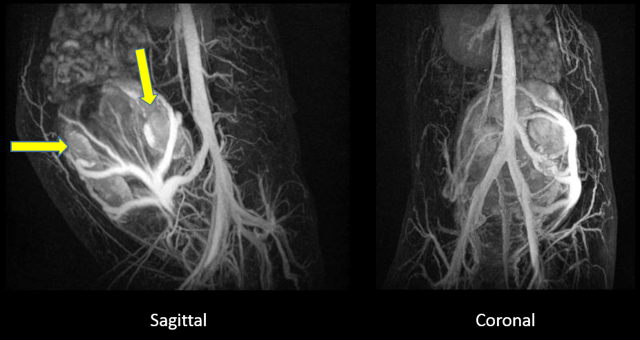
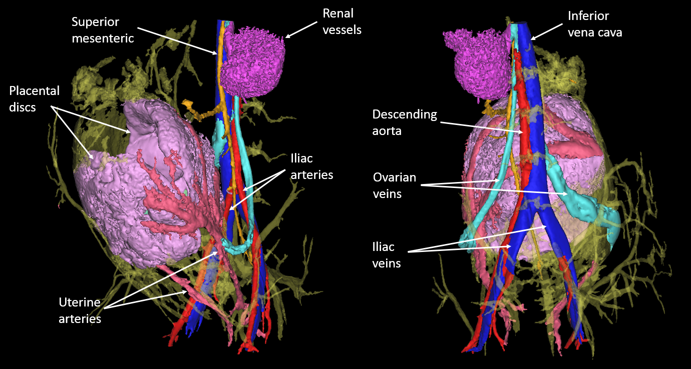
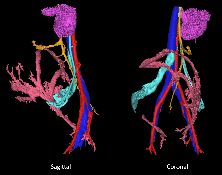
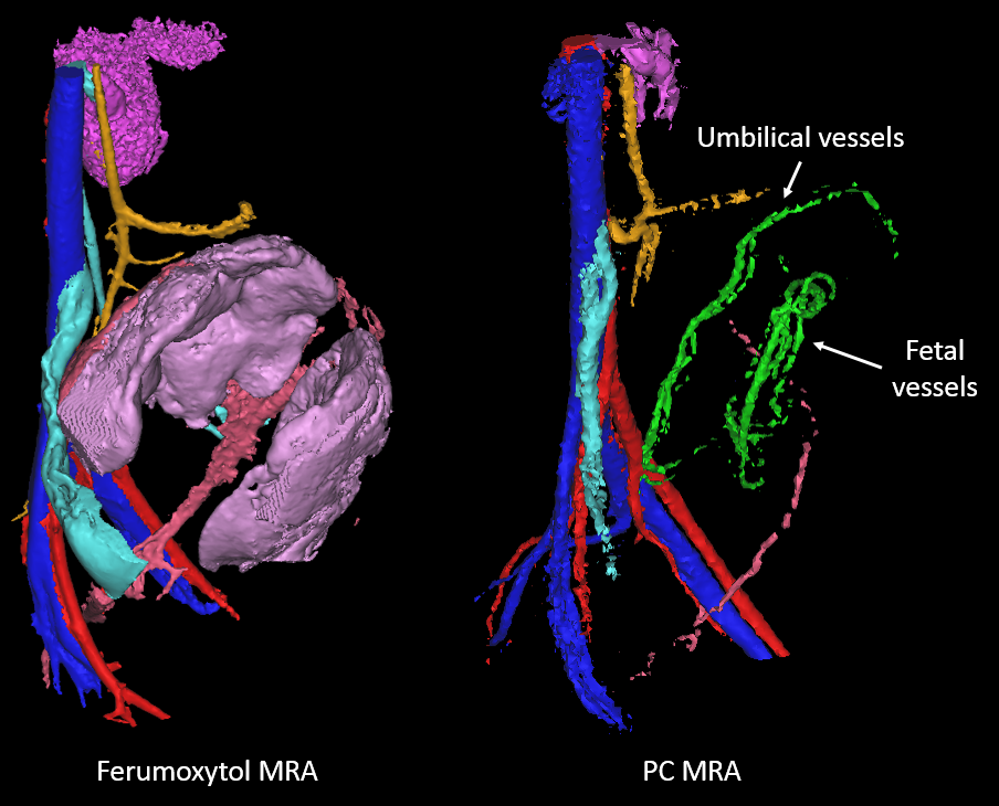
Ferumoxytol-enhanced MRA was successfully reconstructed for two of the monkeys. The third rhesus moved during the post-contrast UTE scan, despite anesthesia, thereby corrupting the image subtraction. Fig. 1 shows sagittal and coronal MIP images of a successful ferumoxytol-enhanced MRA. Fig. 2 shows the segmented volume rendering of the ferumoxytol-enhanced MRA in Rhesus 1, while Fig. 3 shows the same volume rendering with the placenta and smaller vessels removed for clarity. The vessel colors correspond to different vessel networks. The uterine arteries appeared to provide the main blood supply to the placenta, while the ovarian veins provided the main venous return. Fig. 4 shows a comparison of the ferumoxytol-enhanced MRA and PC MRA demonstrating improved depiction of small and slow flowing vessels.Discussion
Ferumoxytol may be a well-suited contrast agent for pregnant women as it is clinically used for iron deficiencies and many pregnant women already require iron supplements. The use of a UTE MRA sequence with ferumoxytol mitigates the substantial T2* shortening effects of ferumoxytol6 and provides a high resolution depiction of arteries, veins, and signal from within the placenta. While we observed excellent image quality in the uterine arteries and ovarian veins, the corresponding uterine veins and ovarian arteries were not detected. This may suggest reduced flow in these vessels relative to their counterparts. Ferumoxytol is a long-lived intravascular agent and in the steady-state it provides the additional benefit of visualizing the maternal blood volume of the placenta, which allows for straightforward volume measurements of the placenta. We did not detect any signal increase in the fetal circulation, suggesting that ferumoxytol does not cross the placenta. However, this also prevents visualization of the fetal vasculature, which is visible with 4D Flow MRI (Fig. 4). As a subtraction technique, this approach is sensitive to misregistration artifacts from motion between the pre and post-contrast acquisition.Conclusion
We demonstrated the feasibility of high resolution, contrast-enhanced MRA with ferumoxytol in a small cohort of healthy, pregnant rhesus macaques. This technique proved to be superior to PC-MRA in detecting utero-placental vessels due to its improved spatial resolution and SNR. Interestingly and importantly, ferumoxytol did not appear to cross the placenta into the fetus.Acknowledgements
The authors acknowledge the support of the NIH Human Placenta Project (NICHD U01HD087216) and NIH grant number P51 OD011106 to the Wisconsin National Primate Research Center. We also thank GE Healthcare for their support.References
1. Macdonald J, Skopos S, Johnson K, Ludwig K, et al. Magnetic Resonance Imaging of Utero-Placental Vascular Flow and Tissue Perfusion in Pregnant Rhesus Macaques. Placenta 2016;85(6): 85.
2. Macdonald J, Skopos S, Johnson K, Francois C, et al. 4D Flow Imaging of the Placenta and Umbilical Cord in the Rhesus Macaque – Initial Experience. 28th Society for Magnetic Resonance Angiography (SMRA). 2016.
3. Webb J, Thomsen H, Morcos S. The use of iodinated and gadolinium contrast media during pregnancy and lactation. European Radiology. 2005;15(6):1234-1240.
4. Johnson KM, Fain SB, Shiebler ML, Nagle S. Optimized 3D Echo Time Pulmonary MRI. MRM. 2013; 70(5): 1241-1250.
5. Johnson KM, Lum DP, Turski PA, Block WF, et al. Improved 3D phase contrast MRI with off-resonance corrected dual echo VIPR. MRM. 2008;60(6):1329-1336.
6. Reeder SB, Smith MR, Hernando D. Mathematical optimization of contrast concentration for T1-weighted spoiled gradient echo imaging. MRM. 2016;75(4):1556-1564.
Figures