4811
4D Flow MRI in the Rhesus Macaque Fetus1Medical Physics, University of Wisconsin - Madison, Madison, WI, United States, 2Comparative Biosciences, University of Wisconsin - Madison, Madison, WI, United States, 3Radiology, University of Wisconsin - Madison, Madison, WI, United States, 4Obstetrics and Gynecology, University of Wisconsin - Madison, Madison, WI, United States, 5Wisconsin National Primate Research Center, University of Wisconsin - Madison, Madison, WI, United States
Synopsis
4D flow measurements were performed with PC-VIPR in rhesus macaque monkeys undergoing healthy pregnancies to determine the feasibility of flow measurements in the fetal vasculature and umbilical cord. Flow measures appeared to be viable for the larger fetal vessels (aorta and IVC), but more variable in smaller vessels with slower flow (umbilical vessels). Image quality improved for later gestational ages as a result of increased vessel area.
Purpose
Assessment of blood flow to the placenta and to and from the fetus is of clinical and research interest to evaluate fetal well-being. We recently demonstrated the feasibility of 4D flow MRI for the assessment of flow in the utero-placental vessels of the pregnant rhesus macaque1,2 In this pilot study, we investigated the feasibility of 4D flow MRI for vessel visualization and the integrity of subsequent quantitative flow measures in the rhesus macaque fetus.Methods
Eight healthy, pregnant rhesus macaques were imaged in a right-lateral position on a 3.0T scanner (Discovery MR750, GE Healthcare) with a 32-channel cardiac or torso coil. All procedures were approved by our institution’s animal care and use committee (IACUC). The gestational ages of the monkeys ranged from early 2nd trimester to early 3rd trimester. 4D flow imaging of the abdomen was performed with a radially-undersampled sequence (PC-VIPR3, 5-point velocity encoding, TR/TE=6.1/2.6ms; FA=8°; VENC=60cm/s; FOV=16x16x16cm3; matrix=192x192x192; isotropic voxel size=0.83 mm, scan duration=610s). All animals were sedated with isoflurane during imaging. Retrospective respiratory gating was used to include only data during expiration (50% efficiency) for image reconstruction. A time-averaged PC angiogram (PC MRA) was generated by combining the magnitude and velocity data similar to complex difference processing. Segmentation of the umbilical cord and fetal vessels was performed on PC MRA images with MIMICS (Materialize). Time-averaged flow was measured in the ascending aorta and inferior vena cava (IVC) of the fetus and the proximal and distal ends of the umbilical cord within EnSight (CEI Inc.) by integrating through-plane velocity components over the segmented vessel areas. A conservation of mass approach was used to assess the internal consistency of PC-VIPR measures by comparing flow in the ascending aorta against the IVC, and flow in the umbilical vessels at both ends of the umbilical cord.Results
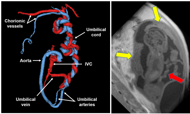
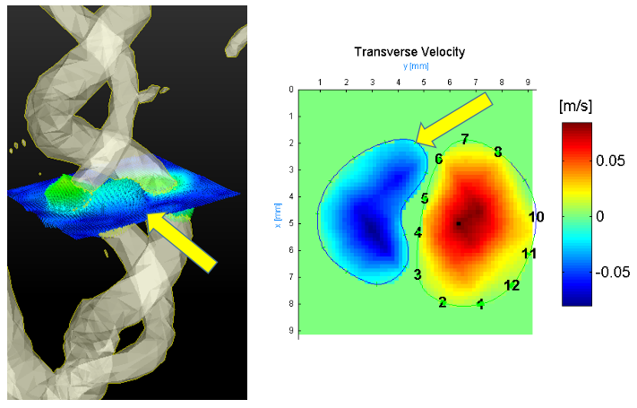
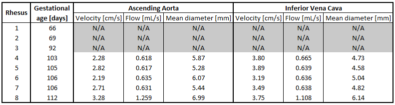
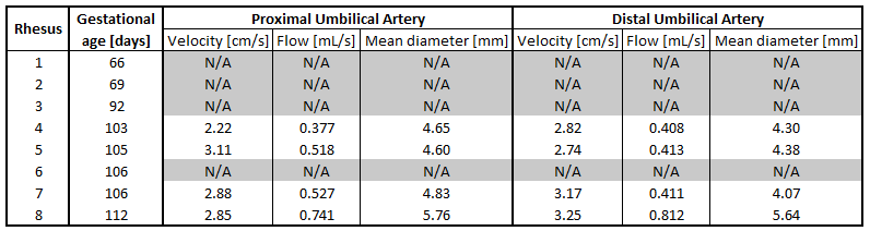
Vessel visualization and flow measurements in the fetal aorta and IVC were feasible in five of the monkeys, while measurements in the umbilical cord were possible in four. The three animals that did not have sufficient SNR for detection of the fetal vasculature were all early-to-mid 2nd trimester. Fig. 1 shows the segmented umbilical cord and fetal vasculature in one rhesus. In all monkeys, only one of two umbilical arteries was visualized within the umbilical cord (Fig 2). Fig. 3 shows a comparison of flow, velocity, and diameter measures in the ascending aorta and IVC for each rhesus. The largest difference in flow between these two vessels was only 12%. Figures 4 and 5 list mean velocity, flow, and mean diameter measures in the umbilical artery and vein, respectively. These measurements demonstrated less consistency, with an average change in flow of approximately 20% between the two ends of the vessels.Discussion
Our pilot study shows that 4D flow acquisitions in fetal and umbilical vessels in the macaque are feasible in the later stages of gestation, from late 2nd trimester onwards. Vessel area and flow were likely not high enough at earlier stages to provide sufficient SNR to resolve vessels above the noise floor. Even in early 3rd trimester, smaller vessels such as the second umbilical artery were not separately resolved in the umbilical cord. Limits in spatial resolution are likely the source of these difficulties, as measurements in resolved vessels showed mean vessel diameters (approx. 4-5 mm) not much larger than our spatial resolution (0.83mm). The fetal aorta and IVC showed internal consistency of flow measurements, suggesting PC-VIPR is a viable tool for measurements in these vessels. Measurements in the umbilical vessels, however, were less reliable and not possible in four monkeys. Partial-volume effects were likely enhanced in these vessels, as a larger fraction of the voxels covering the vessel would have been affected given the smaller vessel diameter. The sedation of the mother also sedated the fetus, which was likely an important factor in the image quality of these acquisitions, as it eliminated fetal motion during scanning. The challenge of fetal motion will need to be addressed in future studies with human subjects who are not sedated.Conclusion
This feasibility study suggests that high resolution 4D Flow MRI acquisitions using PC-VIPR are feasible in the aorta and IVC of the rhesus macaque fetus from late 2nd trimester onwards. Assessment of the umbilical cord is more challenging and might require improved spatial resolution. Image quality likely benefited from the sedation of the mother and fetus.Acknowledgements
The authors acknowledge the support of the NIH Human Placenta Project (NICHD U01HD087216) and NIH grant number P51 OD011106 to the Wisconsin National Primate Research Center. We also thank GE Healthcare for their support.References
1. Macdonald J, Skopos S, Johnson K, Ludwig K, et al. Magnetic Resonance Imaging of Utero-Placental Vascular Flow and Tissue Perfusion in Pregnant Rhesus Macaques. Placenta 2016;85(6): 85.
2. Macdonald J, Skopos S, Johnson K, Francois C, et al. 4D Flow Imaging of the Placenta and Umbilical Cord in the Rhesus Macaque – Initial Experience. 28th Society for Magnetic Resonance Angiography (SMRA). 2016.
3. Johnson KM, Lum DP, Turski PA, Block WF, et al. Improved 3D phase contrast MRI with off-resonance corrected dual echo VIPR. MRM. 2008;60(6):1329-1336.
Figures