4808
Pelvic Floor Structural Alterations of Primipara with Stress Urinary Incontinence After Vaginal Delivery:A MRI Study1Department of Radiology, Tianjin First Center Hospital, Tianjin, People's Republic of China, 2Philips Healthcare, Beijing, China, beijing, People's Republic of China
Synopsis
Vaginal childbirth women have an increasing incidence of stress urinary incontinence(SUI). There are few studies on pathogenesis of SUI and the relationship between the SUI and pelvic floor structure changes. In this study, static and dynamic MRI imaging are performed to describe and assess pelvic floor structure changes in patients with stress urinary incontinence. The results showed that pelvic floor structures changed significantly in the primipara suffering from SUI after vaginal delivery, which suggest a series of pathological state.
Purpose
Stress urinary incontinence (SUI) is a kind of pelvic floor dysfunction disease, which is defined as involuntary leakage of urine when a sudden increase in abdominal pressure. Vaginal delivery is known to be the most important risk factors of SUI[1]. Recurrently, the pathogenesis of SUI is unclear because Levator ani muscle injury, urethral ligament dysfunction, urethral sphincter weakening or other factors may lead to the occurrence of SUI[2]. MRI has the advantage of high soft tissue resolution, which can display the structure of the pelvic floor in detail. In this study, static and dynamic MRI are applied to observe and assess pelvic floor structure changes in patients with stress urinary incontinence.Methods and methods
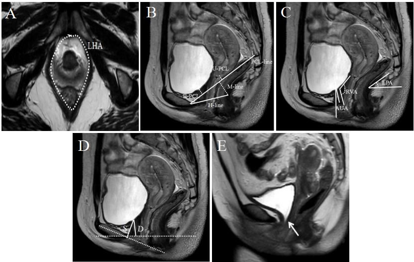
In this prospective study, nineteen vaginally primiparous women with stress urinary incontinence were included as the SUI group.Wwenty-five normal primiparous women were included as the delivery normal(DN) group and seventeen nulliparous were included as the normal control(NC)group. All the subjects underwent static and dynamic MRI in both train and rest state with a 3T MRI scanner (Ingenia, Philips Healthcare, the Netherlands). Two radiologists evaluated and measured the parameters of pelvic structures, including LHA,H line,M line,B-PCL,U-PCL,RVA,AUA,LPA,D,S (as defined in Fig.1) and evaluated whether there was a bladder funnel. The measurements consistency between the two observers were evaluated by intra-class correlation coefficient (ICC). One-way ANOVA was used to compare the measurements difference among three groups. Besides, the different proportion of bladder funnel in the three groups were compared by chi-square test.Results
The consistency between two observers were good(ICC>0.75). In strain state, the measurements including LHA,H line,M line,B-PCL,U-PCL,RVA,AUA,LPA,D,S were different among three groups(P<0.05). And there were significant differences between SUI group and DN as well as between SUI group and NC groups . In the rest state, there were also significant differences among three groups. The measurements of LHA,H line,M line,RVA,AUA,LPA in SUI group were bigger than those in NC group(P<0.05) and B-PCL,U-PCL,D,S were smaller than those of NC group(P<0.05). The proportion of bladder funnel in three group were 78.95% for SUI group), 4% for DN group)and 0% for NC group respectively, and there were significant differences(χ2=39.718, P<0.001).Discussion
The SUI group shows obvious increases of LHA, H line, M line, RVA, AUA and LPA and significant diminutions of B-PCL, U-PCL, D, S, which indicates some degree of levator ani muscle dysfunction, urethral activity increasing, internal urethral orifice with a poor closed integrity and urethral sphincter disability.Conclusion
Pelvic floor structures changed significantly in the primipara suffering from SUI after vaginal delivery, which suggest a series of pathological state. As an auxiliary examination technique, MRI can evaluate the degree and range of the lesions comprehensively and has an excellent clinical application value.Acknowledgements
No acknowledgement found.References
1. Tahtinen RM, Cartwright R, Tsui JF, et al. Long-term Impact of Mode of Delivery on Stress Urinary Incontinence and Urgency Urinary Incontinence: A Systematic Review and Meta-analysis[J]. Eur Urol, 2016, 70(1): 148-158.
2. Pontbriand-Drolet S, Tang A, Madill SJ, et al. Differences in pelvic floor morphology between continent, stress urinary incontinent, and mixed urinary incontinent elderly women: An MRI study[J]. Neurourol Urodyn, 2016, 35(4): 515-521.
Figures