4805
Full 3D high-resolution BOLD imaging of the human placenta with prospective navigation and 2D spatially selective excitation1Radiology and Imaging Sciences, University of Utah, Cottonwood Heights, UT, United States, 2Oregon Health & Science University, 3Department of Obstetrics and Gynecology, University of Utah
Synopsis
Full 3D high-resolution human placenta BOLD imaging at 3T was performed with a free-breathing prospectively navigated sequence using 2D spatially selective excitation. Advantages of this sequence over conventional multi-slice breath hold BOLD include increased SNR, no breath hold recovery periods leading to better time efficiency, and elimination of artifacts from respiratory and fetal motion. T2* maps clearly show the cotyledon architecture of the human placenta.
Purpose
To develop robust novel methods of BOLD MRI for functional imaging of the human placenta.Introduction
MRI has been used for many years for anatomic evaluation of the placenta. More recently, BOLD MRI seeks to move beyond anatomic depiction to provide functional information about the placenta. Gadolinium based contrast agents have been used in animal models to depict vascular structures in the placenta and to quantify blood flow (1) but are not yet considered safe for human placenta imaging (though recent nonhuman primate research suggests they may be (2)). T2* measured with non-contrast placental BOLD MRI provides an endogenous contrast mechanism that reflects deoxyhemoglobin concentration. Features on placental T2* maps have been shown to correlate with placental vascular anatomy and perfusion depicted with DCE-MRI in macaque placentas (3). BOLD MRI of the placenta offers the promise of non-invasive quantification of vital placental oxygen delivery to the developing fetus. Several challenges exist in BOLD MRI of the human placenta. The placenta moves with maternal breathing, requiring BOLD to be performed during multiple breath holds. Fetal motion causes artifacts. The location and shape of the placenta are variable. We have addressed these limitations with a novel prospectively navigated free-breathing sequence (FB-BOLD) which utilizes two-dimensional volume-selective excitation to perform high resolution 3D BOLD MRI of the entire human placenta without respiratory or fetal motion artifact.Methods
The FB- BOLD sequence was used to image a healthy pregnant volunteer at 38 weeks gestation, after IRB approval and informed consent. An imaging field of view fitted to the placenta was prescribed in an oblique coronal plane, excluding most of the gravid uterus and fetus. The pulse sequence designs a 2D excitation tailored to select spins within the prescribed imaging volume. This design process takes less than 1 second and does not perceptibly increase sequence setup time. The FB-BOLD sequence acquires a navigator with a 1cm diameter pencil-beam excitation oriented along the frequency direction of the imaging volume at the beginning of each sequence repetition. This is analyzed in real time to sort the data acquisitions into different bins based on respiratory motion. Once the current bin has been determined, feedback to the running sequence occurs in real-time to set the appropriate next phase encode step for that bin. Excitation is then performed with the 2D excitation which excites the imaging volume but nothing outside of it. Multi-echo data acquisition is then performed with centric Cartesian k-space ordering. Eventually, one bin contains a complete k-space and the sequence is complete. Sequence parameters for the FB-BOLD sequence include TR = 150ms, TE = (5, 12, 19, 26, 33, 40, 47, 54, 61, 68, 75ms), isotropic resolution of 2mm, matrix size 128 x 88 x 36, FOV 250 x 171 x 72 mm, total image time 14:10, flip angle 10, pixel bandwith 150Hz, efficiency (i.e. fraction of data used in final image) 59.4%. Imaging was performed on a 3T Siemens Trio system.
Results
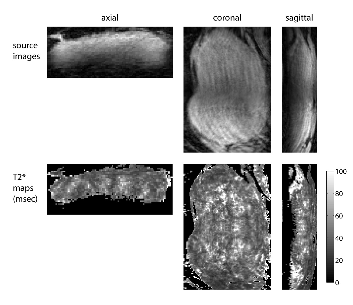
Figure 2 shows an axial T2-weighted HASTE image of the placenta, showing the approximate prescribed profile of the 2D excitation. Figure 4 shows the results of the free-breathing sequence, including T2* maps of the placenta.Discussion
We have demonstrated the feasibility of a prospectively gated free-breathing placental BOLD sequence with spatially selective 2D excitation (FB-BOLD) which can acquire full 3D high-resolution T2* maps of the human placenta. This method is applied to placenta imaging for the first time; we have previously demonstrated a similar technique for free-breathing renal BOLD (4). Because the FB-BOLD sequence uses 3D acquisition, it gives source images with much higher SNR than conventional multi-slice multi-breath hold BOLD, leading to more accurate T2* maps. Artifacts from fetal motion or motion of other structures outside the imaging field of view are eliminated by the volume-selective excitation. Some motion artifact is seen in the coronal plane images representing motion in the right-left direction which was not captured by the superior-inferior directed navigator. This artifact might be eliminated by additional navigator directions, which is a subject for future investigation. The time efficiency of the FS-BOLD sequence (59.4% in this example) is better than conventional multi-slice breath hold methods, since recovery time is necessary between breath holds which typically results in less than 50% efficiency for conventional multi-slice BOLD.Conclusion
A novel prospectively gated free-breathing placental BOLD sequence with volume selective 2D excitation can acquire full 3D high resolution T2* maps of the human placenta free of respiratory and fetal motion artifacts.Acknowledgements
The authors gratefully acknowledge support from 2R01DK063183-06 (NIDDK) and 1U01HD087182-01 (NICHD).References
1. Frias AE et al: Using dynamic contrast-enhanced MRI to quantitatively characterize maternal vascular organization in the primate placenta. Magn Reson Med 73: 1570-1578, 2015.
2. Oh KY, et al: Gadolinium Chelate Contrast Material in Pregnancy: Fetal Biodistribution in the Nonhuman Primate. Radiology, 276: 110-118, 2015.
3. Schabel MC et al: Functional imaging of the nonhuman primate Placenta with endogenous blood oxygen level-dependent contrast. Magn Reson Med 76: 1551-1562, 2016.
4. Morrell G, Kaggie J, Lee VS: Full 3D Renal BOLD MRI in
Clinically Realistic Scan Times with 2D Volume Selective Excitation. 23rd
Meeting ISMRM, Toronto, 464, 2015.
Figures
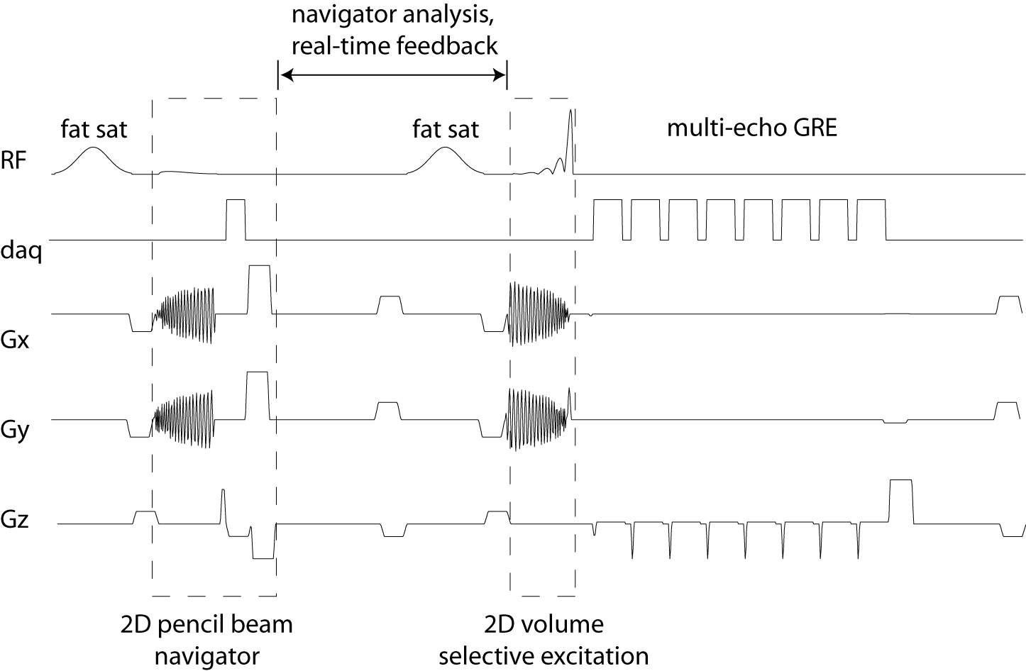
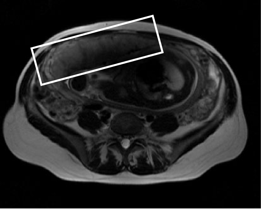
Figure 2: Axial T2-weighted HASTE image showing the placenta, with the cross-section of the imaging volume of the FB-BOLD sequence shown by the white rectangle overlay. The FB-BOLD volume includes the entire placenta but excludes other structures such as the fetus.