4802
Evaluation of Placenta Motion throughout GestationThomas Martin1,2, Dapeng Liu2, Teresa Chanlaw3, Sherin U. Devaskar3, Carla Janzen4, Tess Armstrong1,2, Yutaka Natsuaki5, Daniel Margolis2, Rinat Masamed2, Holden Wu1,2, and Kyunghyun Sung1,2
1Biomedical Physics, University of California, Los Angeles, Los Angeles, CA, United States, 2Radiological Sciences, University of California, Los Angeles, Los Angeles, CA, United States, 3Department of Pediatrics, University of California, Los Angeles, Los Angeles, CA, United States, 4Department of Obstetrics and Gynecology, University of California, Los Angeles, Los Angeles, CA, United States, 5Siemens Healthcare, Los Angeles, CA, United States
Synopsis
Proper placental function is essential for normal fetal development. MRI can easily assess the growth and development of the placenta throughout gestation due to its capabilities in functional imaging. However, uterine contractions, and fetal and maternal motion can lead to inaccuracies in the quantitative assessments. In this we assessed the impact of extraneous motion on placental imaging by using a 3D multi-echo golden angle radial sequence to generated dynamic images of the placenta.
Introduction
Proper placental function is essential for normal fetal development. The placenta is known to transfer nutrients to the fetus with its high blood volume1. MRI can easily assess the growth and development of the placenta throughout gestation due to its capabilities in functional imaging2. However, uterine contractions, and fetal and maternal motion can lead to inaccuracies in the quantitative assessments. It is not well documented on how much these motion characteristics weigh in and need attention in the final assessment. In this study we aim to assess the impact of extraneous motion on placental imaging by using a 3D multi-echo golden angle radial sequence to generated dynamic images of the placenta.Methods
To assess the effect of motion on the placenta 7 healthy pregnant subjects were scanned at two different times in their gestation (first scan 14-16 weeks, second scan 19-22 weeks) for a total of 14 scans on a 3T scanner (Prisma, Siemens Healthcare, Erlangen, Germany). A 3D multi-echo golden angle (GA) radial GRE sequence was used to acquire the raw data, which was then reconstructed offline using k-space weighted image contrast (KWIC)3 to create high spatial resolution dynamic image series without much streaking. There were two main protocols used for imaging and are listed in table 1. The multi-echo imaging was used for R2* quantification, but only the first echo that imparted the highest signal was used for reconstruction. Motion was semi-quantitatively assessed by assessing the absolute difference between the first image of the timed series and all other subsequent images in the time frame of scanning (see figure 1). Two regions-of-interests (ROI) were drawn to segment the placenta and the outer uterus on three orthogonal slices. A T2-HASTE image was used as reference for the placental location. To reduce the effect of noise and streaking artifacts the subtraction image was filtered using a window leveling so that majority of the remaining non-zero pixels correlated with motion. The number of pixels were then counted and normalized to derive the percent change in the # of pixels across time and then averaged across the entire scan time frame.Results
Figure 1b shows that there is visible motion between the first and last images of the dynamic series of a subject at 21 weeks gestation, and figure 1c shows that the motion timed series is in agreement with the subtraction image. The sharp rise near t = 50s is due to noise build up between the reference time point and later timed images, and not necessarily motion. Figure 2 shows a different subject at 21 weeks in whom the uterus expands and then contracts during the middle of the scan across a 2min interval. It shows that the motion is not abrupt, but smooth over a long period of time. Figure 3 shows the mean percent change of pixels for the total scan time (4-6min) as well as half of the total scan time (2-3min). There is a noticeable decrease in the mean percent pixel change for scan time of 2-3min compared to longer scan time. There is also an overall slight trend of decreased motion throughout the scan as the gestation weeks increase. The largest amount of pixel change was about 22%.Discussion
In this study we have shown a semi-quantitative analysis of uterine and placental motion throughout gestation. The average range across the total scan time of the percent pixel change was about 5-15%. However, due to the noise accumulation there was a sharp rise in the percent pixel change for every case around t=50s. This could be improved by removing the noise bias. Also by reducing the scan time the amount of uterine motion is decreased. However, there was still a significant amount of motion detected that could be problematic in interpretations.Conclusion
We have demonstrated that there is placental and uterine motion in the first half of gestation under long scan times (>4min). Decreasing the scan time can reduce the motion within the scan time. However, for perfusion imaging with multiple scans, motion correction should be considered.Acknowledgements
Funding for project is provided by NICHD U01-HD087221.References
1. Rossant et al., NRG 2001
2. P. Gowland, Seminars in Fetal & Neonatal Medicine 2005
3. Song et al., MRM 2004
Figures
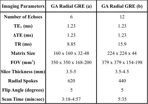
Table 1:
Representative sequence parameters for the in vivo placenta scans.
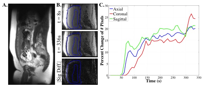
Figure
1 demonstrates how
the motion estimation is evaluated. A is the reference image to identify placenta
location. To identify the motion the absolute difference between the first time
point and the other time points is performed (B). The ROI’s are used to create a mask to reduce
noise. C plots the
percent change of the number of pixels within the ROI’s.
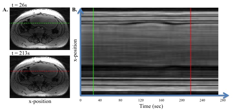
Figure
2 Shows the motion
of the uterus at the cross section of axial slices (A) identified by the green and red lines. B shows that the
uterus expands and contracts during the middle of the scan.
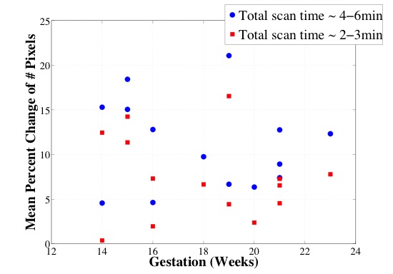
Figure
3 shows the mean
percent change of the number of pixels throughout gestation for scan times of
4-6min and 2-3min. There is a decrease
in the mean percent change of pixels when the scan time is decreased. There is
an overall trend of decreased motion as gestation time increases.