4787
Evaluation of prostate cancer on diffusion weighted imaging; Can FOCUS and synthetic diffusion weighted imaging with FOCUS contribute to the Prostate Imaging Reporting and Data System (PI-RADS) version 2.0?Motoyuki Katayama1, Takayuki Masui1, Kei Tsukamoto1, Mitsuteru Tsuchiya1, Masako Sasaki1, Yuki Hayashi1, Takahiro Yamada1, Mitsuharu Miyoshi2, Tetsuya Wakayama2, and Harumi Sakahara3
1Radiology, Seirei Hamamatsu General Hospital, Hamamatsu, Japan, 2GE Healthcare Japan, Japan, 3Radiology, Hamamatsu University School of Medicine, Hamamatsu, Japan
Synopsis
We
evaluated 47 patients suspected of having prostate cancers on synthetic DWI
calculated from FOCUS DWI with PI-RADS version 2.0. Compared with conventional
FOV DWI, FOCUS DWI is more useful for evaluation of prostate cancer with high
spatial resolution and less distortion. S-DWI is able to enhance diagnostic
ability of FOCUS without image degradation, which might be one of the best
combinations, and contribute to the Prostate Imaging Reporting and Data System (PI-RADS)
version 2.0.
Introduction
MR imaging has played an important role in evaluating prostate lesions. Especially, diffusion weighted imaging (DWI) with single-shot echo planar imaging (ss-EPI) is a useful tool for detection of the prostate cancer. However, DWI with ss-EPI is susceptible to inhomogeneity of the static magnetic field and may result in image distortion. With recently introduced Field-of-view (FOV) optimized and constrained undistorted single-shot (FOCUS, GEHC), we can decrease the required readout duration for single shot EPI by using a 2D spatially selective echo-planar RF excitation pulse and a 180 degrees refocusing pulse, consequently, and can acquire high spatial resolution images with less distortion (Figure.1). Synthetic DW Imaging (s-DWI) is a new method that can calculate DWIs for any b-value from at least two DWIs using different b-values. On the other hand, the revised version of the Prostate Imaging Reporting and Data System (PI-RADS) has been popular for the assessment of the prostate cancer.Purpose
The purpose of this study is to compare delineation of prostate cancers in FOCUS DWI and Synthetic (S)-FOCUS DWI with those in conventional FOV DWI by means of PIRADS version 2.0.Materials and Methods
47 male patients (mean age: 67.6 years old, ranged from 50-78 years old), suspected of having cancers in the peripheral zone of the prostate glands, were included in this study, who underwent MRI on a 3T unit (MR750, MR750W, GE Healthcare) before or after biopsy. The parameters of FOCUS DWI were as follows; FOV: 24*12cm, Matrix: 128*64, section thickness: 4 mm, TR/ TE: 4000 /55 msec and those of conventional DWI were as follows; FOV: 32*25 cm, Matrix 128*96, section thickness: 4 mm, TR/ TE: 4000/ 63-66 msec, respectively. The b-values of FOCUS imaging were 0 and 800, and those of conventional DWI were 0 and 1500. S-FOCUS DWI with b-value of 1500 calculated from FOCUS with those of 0 and 800. Apparent diffusion coefficient (ADC) of the tumor in each imaging was calculated with FUNCTOOL (GE Healthcare). The scores (1, 2, 3, and 4 including 5) on PIRADS assessment of DWI with or without ADC maps were applied to the tumor detection on each imaging. The contrasts of the signal intensity (SI) of the tumor against that of the prostate tissue adjacent to the tumor in each imaging were measured on PACS system.Results
Clinical stage of the prostate cancer were as follows; negative: n =10, ≦T2: n = 34, T3: n = 1 (T3a), and T4 : n = 2 (bone metastases), respectively. Maximum Gleason scores (GS) of the tumor were as follows; no cancer: n = 10, GS 6: n = 11, GS 7 (3+4): n = 14, GS 7 (4+3): n = 4, GS 8≦ n = 8, respectively. The total prostectomy were performed in 12 patients. Correlation Coefficient (R) of calculated ADC of tumor between in FOCUS imaging and in Conventional imaging was 0.87. Overall image quality, delineation of tumor, image distortion, blur, and motion artifacts in FOCUS DWI and S-FOCUS DWI were superior to those in C-DWI, respectively (figure 2). The contrast of tumor against the peripheral tissue in S-FUCUS DWI was superior to that in C-DWI and FOCUS-DWI (p<0.017) (figure 3). Figure 4 shows the table of the detectability of each imaging wit PIRADS version 2.0. The accuracy on S-FOCUS DWI with ADC map was the best of all. Figure 5 shows the each imaging.Conclusion
It is useful to use FOCUS for evaluation of prostate cancer with high spatial resolution and less distortion. S-DWI is able to enhance diagnostic ability of FOCUS without degradation of image quality, and can contribute to the Prostate Imaging Reporting and Data System (PI-RADS) version 2.0. S-FOCUS DWI can be obtained to have high b-value without prolongation of imaging time and loss of the signal intensity.Acknowledgements
No acknowledgement found.References
Saritas EU, Cunn ingham CH, et al. Magn Reson Med. 200 8 Aug; 60(2): 468-73Figures
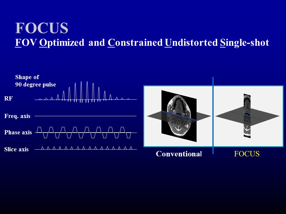
Scheme
of FOCUS: Single shot EPI by using a 2D spatially selective echo-planar RF
excitation pulse and a 180 degrees refocusing pulse, consequently, acquire high
spatial resolution images with less distortion.
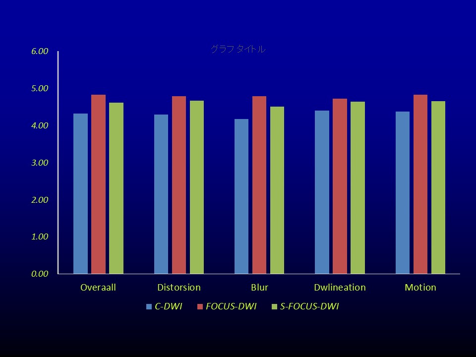
Qualitative
analyses of each imaging.
Overall
image quality, delineation of tumor, image distortion, blur, and motion artifacts
in FOCUS DWI and S-FOCUS DWI were superior to those in C-DWI, respectively.
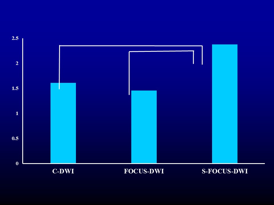
SI tumor / SI peripheral tissue. SI tumor / SI peripheral tissue in S-FOCUS
DWI was superior to that in FOCUS.
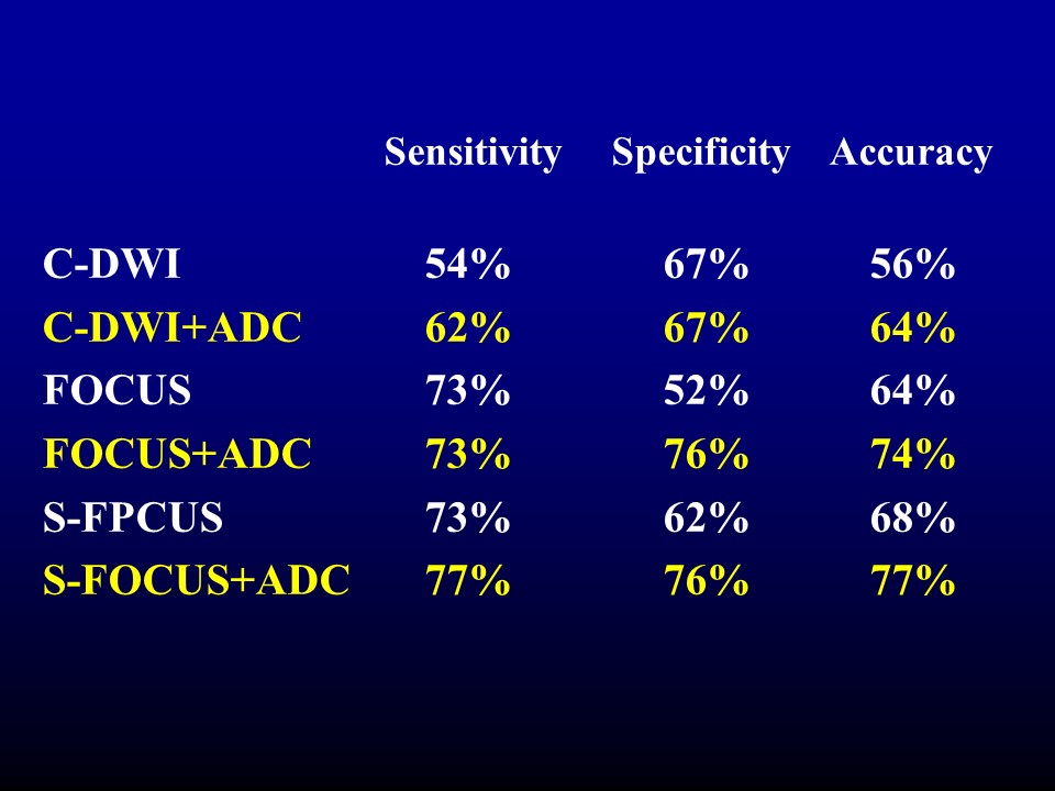
Table of the detectability of each imaging with the tumor with PIRADS
version 2.0.
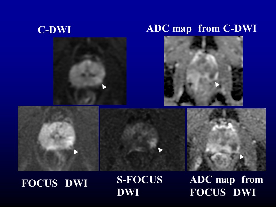
The images of 68-year old man with prostate tumor
(Gleason score7=3+4) in the left peripheral zone (arrow head).