4775
Evaluation of three-dimensional magnetic resonance imaging of autopsied human heart specimens for computational modeling of congenital heart diseases1Department of Radiology, National Cerebral and Cardiovascular Center, Suita, Osaka, Japan, 2Division of Medical informatics, National Cerebral and Cardiovascular Center, Suita, Osaka, Japan, 3Department of Pathology, National Cerebral and Cardiovascular Center, Suita, Osaka, Japan
Synopsis
This study aimed to compare the visibility of a formalin-fixed heart using various 3D MRI sequences as well as to determine the optimal sequence for computational modeling of congenital heart diseases. Our results demonstrated that MPRAGE showed the best contrast with good image quality for imaging of the myocardium and the vascular structure when surrounded by normal saline in a plastic container. We believe that computational cardiac modeling of human autopsied heart specimens using MPRAGE plays a critical role in education and/or research.
Introduction
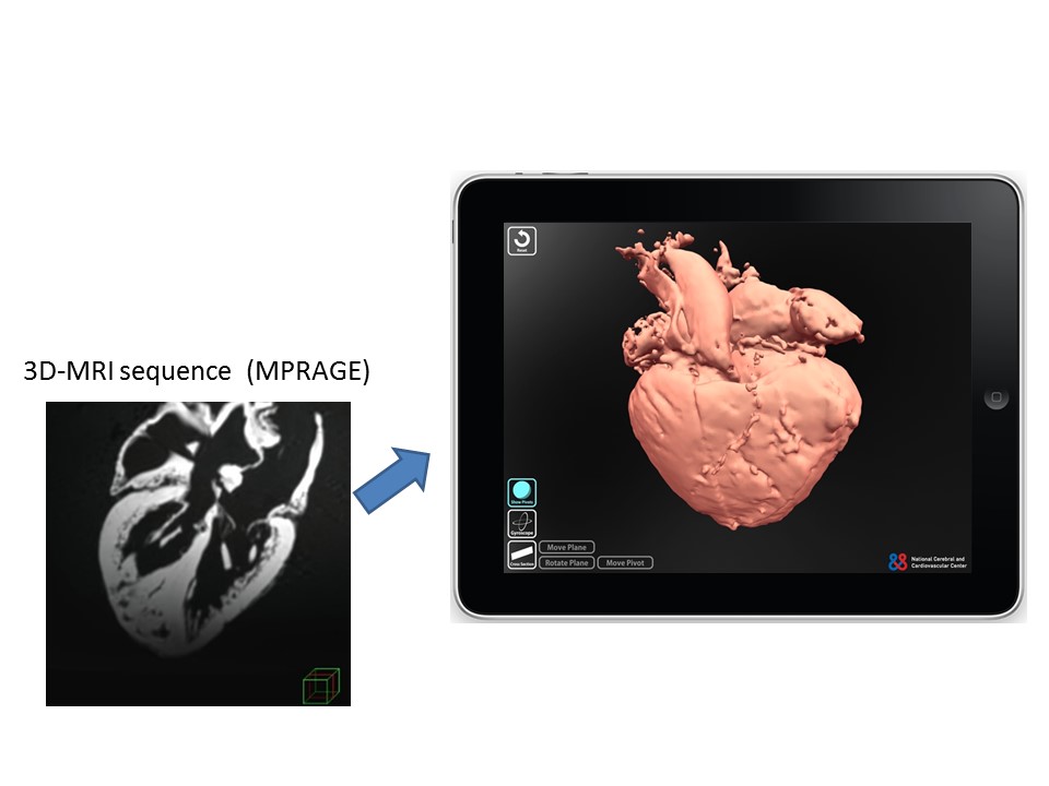
Autopsied human heart specimens with various types of congenital heart diseases are valuable for medical education and/or surgical simulation because they aid in the understanding of the complex anatomical structure of the cardiovascular system. Due to the decreasing number of autopsies being performed and the deterioration of human heart specimens over time, digitalization of these valuable specimens is an effective measure for capturing this data. Computational cardiac modeling using 3D image data has played a critical role in education and/or research (Figure 1). Because 3D MRI sequences provide good image contrast with high spatial resolution, they are used for clinical cardiac imaging such as whole heart coronary MRA and coronary wall imaging. Only a small number of reports on ex vivo heart imaging by MRI can be found1,2; however, the optimal MRI sequence for the imaging of autopsied human heart specimens fixed by formalin, which replaces water in cardiac tissue, is uncertain.Purpose
This study aimed to compare the visibility of a formalin-fixed heart using various 3D MRI sequences as well as to determine the optimal sequence for computational modeling of congenital heart diseases.Methods
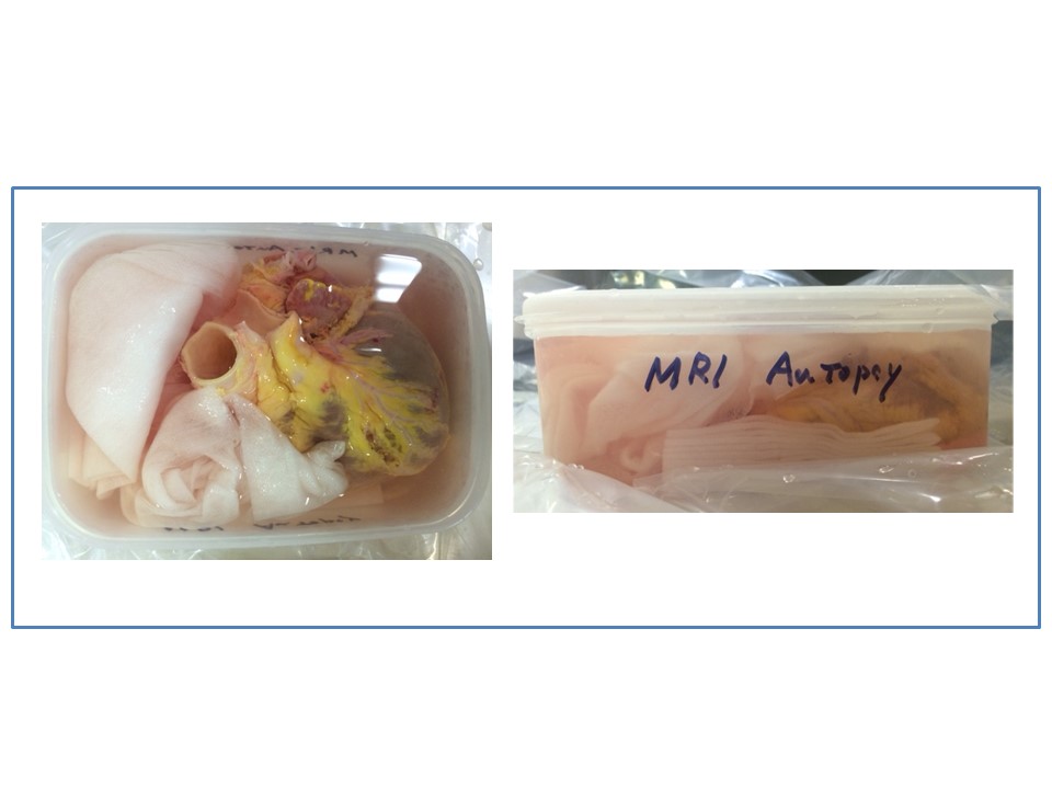
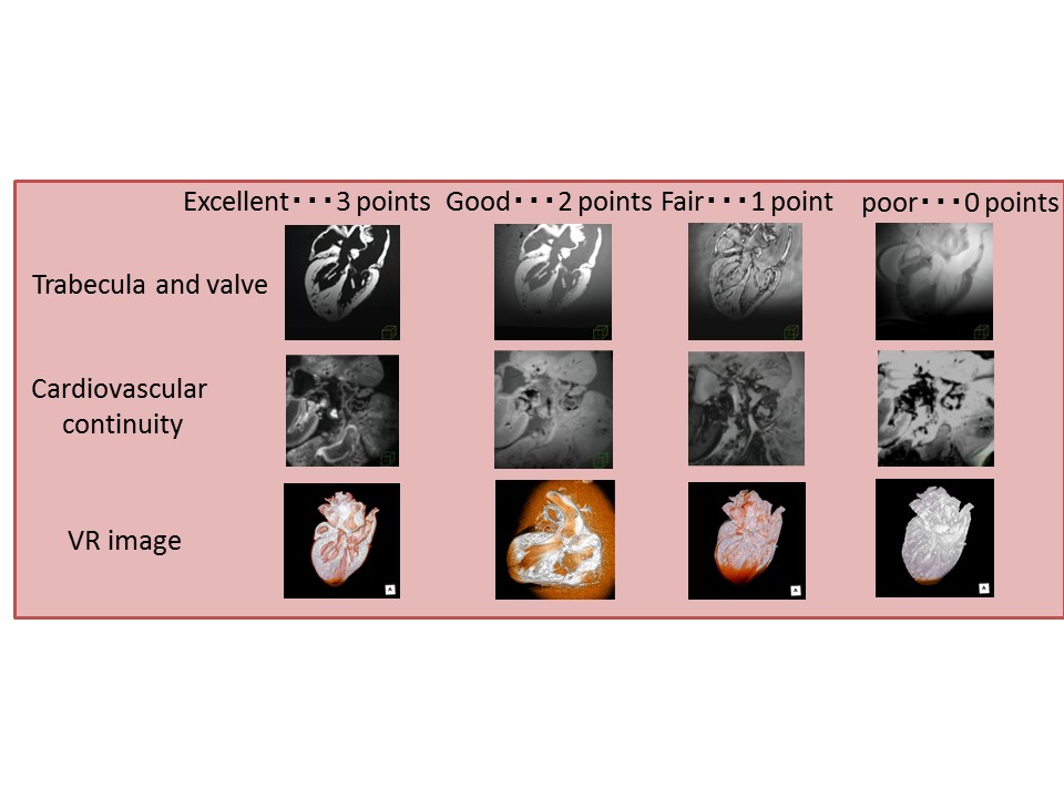
Five human hearts with various types of congenital heart diseases, which were obtained during autopsy and preserved from 1 month to 10 years with formalin fixation, underwent ex vivo MRI with a 3T clinical machine (MAGNETOM Verio; Siemens). Different types of 3D MRI sequences were acquired with the same spatial resolution (voxel size 1.0x1.0x1.0 mm): T2-SPACE (T2-weighted Sampling Perfection with Application optimized Contrasts using different flip angle Evolutions; the 3D-turbo spin echo sequence with a constant flip angle acquires bright blood imaging), True-FISP (True Fast Imaging with Steady state Precession; SSFP imaging), MPRAGE (Magnetization-prepared Rapid Acquisition with Gradient-echo; inversion-recovery-based T1WI), and FLASH (Fast Low Angle Short; basic GRE sequence). The autopsied heart was scanned in a plastic container filled with normal saline (Figure 2). We compared the contrast ratio between the myocardium and the ventricular lumen as well as between the vascular wall and the lumen. In addition, we visually assessed the cardiovascular structure using MPR and 3D images (Figure 3).Results
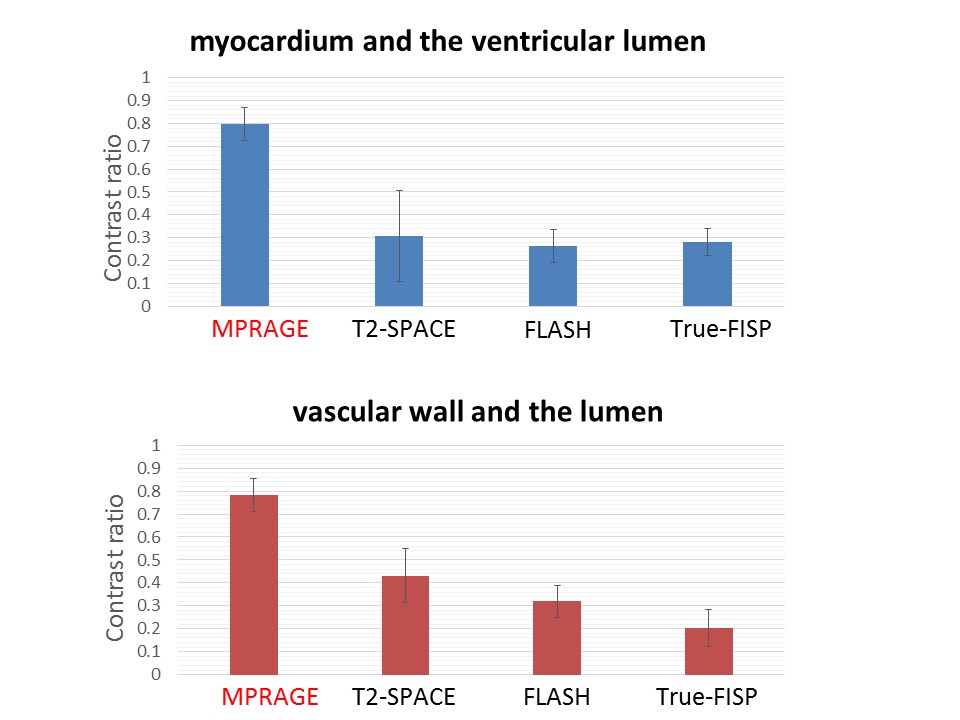
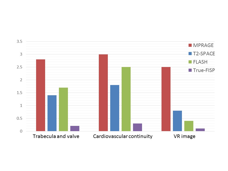
The average contrast ratios between the myocardium and the ventricular lumen in MPRAGE, T2-SPACE, FLASH, and True-FISP were 0.80±0.07, 0.31±0.20, 0.26±0.07, and 0.28±0.06, respectively (Figure 4). The average contrast ratios between the vascular wall and the lumen in MPRAGE, T2-SPACE, FLASH, and True-FISP were 0.78±0.07, 0.43±0.12, 0.32±0.07, and 0.20±0.08, respectively (Figure 4). MPRAGE showed the best contrast for imaging of both the myocardium and the vascular structure. Furthermore, by visual assessment, MPRAGE provided more detailed information regarding the cardiovascular continuity and morphology as compared to other 3D sequences (Figure 5).Discussion
Our study showed that MPRAGE provided the optimal sequence for the imaging of human autopsied heart specimens. MPRAGE is a 3D-inversion recovery-based GRE sequence that offers heavy T1WI3. For imaging of macroscopic specimens, high T1 contrast provided the best contrast between the cardiovascular structure and surrounding normal saline in a plastic container. Further adjustment of the pulse sequence is necessary for more precise visualization of these aspects. The autopsied heart specimens were fixed by formalin, which replaced water in the cardiac tissue. Therefore, the MR signals in the T2 weighted 3D-TSE and SSFP sequences were weak, and contrast ratios decreased.Conclusion
We confirmed that MPRAGE is the optimal sequence for computational modeling of human autopsied heart specimens with congenital heart diseases.Acknowledgements
No acknowledgement found.References
1. Hsu JC et al. Magnetic resonance imaging of chronic myocardial infarcts in formalin-fixed human autopsy hearts. Circulation. 1994 May;89(5):2133-40.
2. Jackowski C et al. Postmortem unenhanced magnetic resonance imaging of myocardial infarction in correlation to histological infarction age characterization. Eur Heart J. 2006 Oct;27(20):2459-67.
3. Brant-Zawadzki M et al. MPRAGE: A three-dimensional, T1-weighted, gradient-echo sequence—initial experience in the brain. Radiology. 1992; 769-775
Figures