4756
Cardiac image acquisition by SENSE combined compressed sensing with three dimensional quantification1Department of Electrical Engineering, Kwangwoon University, Seoul, Korea, Republic of, 2Department of Radiology, Severance Hospital, Yonsei University College of Medicine, Seoul, Korea, Republic of, 3Brain Research Laboratory, Children’s National Health System, Washington, DC, United States
Synopsis
SENSE combined compressed sensing technique is applied to multi-slice cardiac CINE imaging with breath holds. As to the compressed sensing, ITSC is used, which truncates small transformed coefficients in r-f domain to make data sparse, and it restores the measured data in k-t domain iteratively until the reconstructed images converge. Variation of ejection fraction (EF) is measured for two set of experiments, one from regenerated data set by resampling the original data set, and the other from real measurements. Using the variation of EF and normalized mean square error clinical usefulness of the technique is demonstrated.
Purpose
MR cardiac CINE imaging is a useful tool to diagnose cardiac function and anatomy non-invasively. We combined SENSE and compressed sensing technique (ITSC) to increase the level of compression, and investigated the effects of compression on the clinically useful parameters such as ejection fraction with quantitative evaluation.
Methods
ITSC is a compressed sensing technique that truncates small transformed coefficients in the r-f domain to make the data sparse, and it restores the measured data in the k-t domain iteratively until the reconstructed images converge1. Since ITSC does not involve a large-size matrix inversion, it’s fast and is easily combined with other techniques, e.g., SENSE2. We combined ITSC and SENSE to increase the total compression factor3. As to the reconstruction, ITSC reconstruction is performed first to make fold-over images, and then SENSE reconstruction is performed to unfold the images.
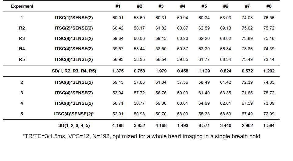
We have performed two set of in-vivo cardiac imaging with volunteers. For the first set of experiments, we have acquired data with a small compression factor (ITSC factor=1, SENSE factor=2), and then various data set (ITSC factor = 2, 3, 4, and 8 with SENSE factor of 2) were regenerated by resampling the data retrospectively. Using the regenerated data set, normalized mean square error (NMSE) and ejection fraction (EF) were evaluated. EF is defined as the ratio of ejection (stroke) volume divided by the end-diastolic volume. For the second set of experiments, we have performed in-vivo cardiac imaging with various ITSC factors of 3, 4, and 8 and SENSE factor of 2. For both set of experiments, balanced SSFP sequence was used with TR/TE=3.88/1.94ms, VPS=8, matrix size (N)=256, and 12 slices of short-axis cardiac CINE images were acquired with a slice thickness of 8mm. For the first set of experiments, one slice CINE images per one breath hold were acquired. In the second set of experiments, 3, 4, 8, and 12 slices CINE images were acquired per a breath hold. To reduce evaluator-dependent variations in the segmentation of blood volume in the left ventricle, a computer program is developed and EF is evaluated automatically with little operator’s effort.
Results
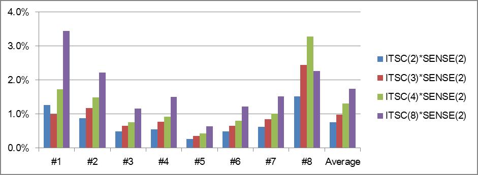
Figure 1 shows NMSE for various compression factors using the regenerated data set. As seen in Fig.1, NMSE with a total compression factor of 8 was less than 1%. NMSE is increasing as compression factor (CF) increases, approximately in proportion to CF0.63.
Table 1 summarizes EF for various compression factors. The upper part of the table is with the data from the first set of experiments. The resampled data were represented as R2~R5. Variation of EF by the compression was measured by the standard deviation. Note that the standard deviation is less than 2%. The lower part of the table is with the data from real measurements as denoted by 2~5. With the second set of experiments, the variation of EF is less than 4.2%. Since each data were measured separately in the second set of experiments, the variation appears larger than that in the first set of experiments where they were from the same data set with only different sampling factors.
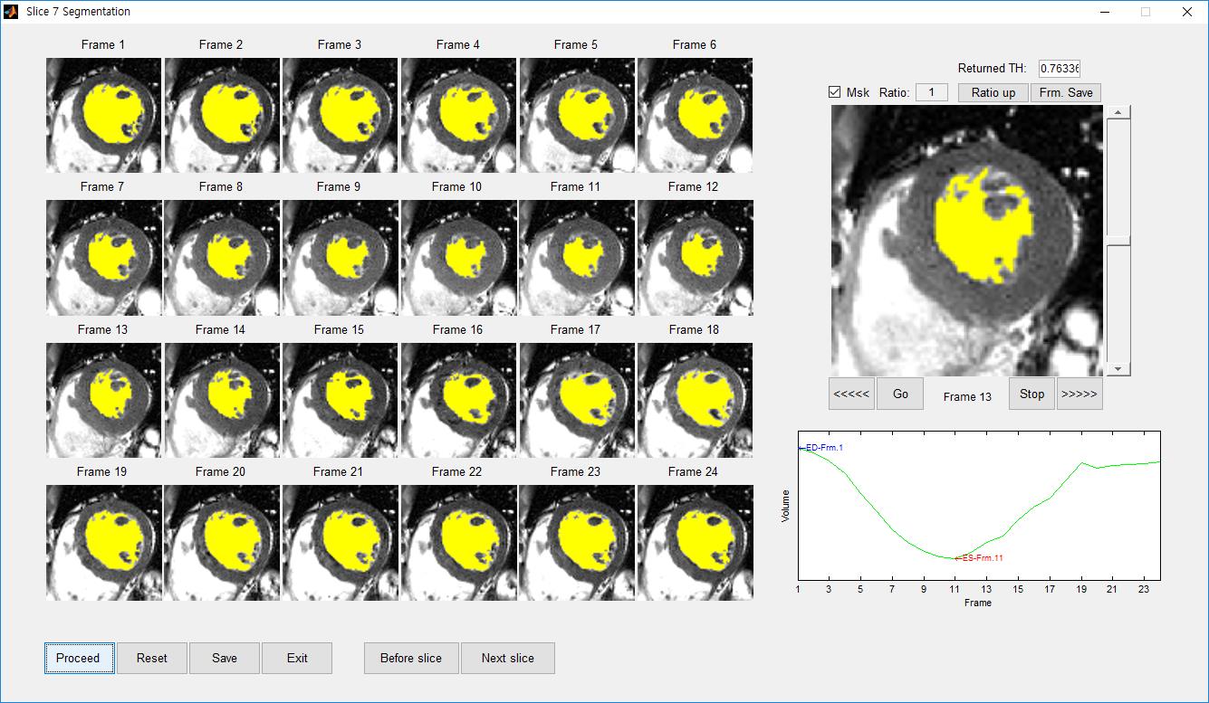
Figure 2 shows a screen of the program to evaluate EF automatically. It displays all the cardiac frames on the screen, and it also provides a window for a CINE display. Segmentation is based on a region growing. Segmented region was displayed in yellow color. The area of the segmented region is displayed in the right bottom window as a function of cardiac cycle.
Discussion
Compressed sensing (x4) with SENSE (x2) provides high quality multi-slice cardiac CINE images within 3 breath-holds. It reduces measurement time, and provides accurate 3-D information by reducing error in the slice registration and motion artifact. EF was reliably obtained from the multi slice cardiac CINE images irrespective of the compression factor. Automatic evaluation of EF reduces evaluator’s efforts substantially, which can be used routinely for clinical diagnosis.Conclusion
The ITSC combined with SENSE was successfully applied to multi-slice cardiac CINE images. NMSE with a total compression factor of 8 was less than 1%. Variation of EF depending on the compression factor was less than 4.2%, thus the compression technique can be useful for clinical applications. Other clinical evaluations are currently under investigation as a function of compression factor.Acknowledgements
This work was supported by the National Research Foundation of Korea (NRF) grant funded by the Korea government (MSIP) (NRF-2015R1A2A2A03005089).References
[1] Park J, Hong HJ, Yang YJ, et al. Fast cardiac CINE MRI by iterative truncation of small transformed coefficients. Investigative Magn Reson Imaging 2015;19(1):19-30.
[2] Pruessmann KP, Weiger M, Scheidegger MB, et al. SENSE: Sensitivity encoding for fast MRI. Magn Reson Med 1999;42(5):952-62.
[3] Park J, Kim PK, Yang YJ et al. Whole heart coverage in a single breath hold using compressed sensing combined with parallel imaging. ISMRM 24th Annual Meeting & Exhibition 2016; Cardiovascular Image Processing 2623, 2016.
Figures