4747
Improved T2-weighted 3D FLAIR from a compact, lightweight 3T scanner with high-performance gradients1Radiology, Mayo Clinic, Rochester, MN, United States, 2MRI, GE Global Research, Niskayuna, NY, United States
Synopsis
A compact, low-cryogen 3T MRI scanner has been developed employing high-performance gradients capable of simultaneously achieving 80 mT/m and 700 T/m/s. A comparison study of T2-weighted 3D FLAIR in 16 clinical patients graded by two neuroradiologists has been performed. The compact 3T system performed equally well to a standard whole-body system in terms of motion artifacts and cerebellar folia conspicuity, and performed better in terms of signal-to-noise ratio, lesion conspicuity, gray/white contrast, and overall exam quality.
Purpose
The purpose of this NIH-funded initiative was to design, build, and evaluate a high-performance, low-cryogen, compact 3T MRI system for with gradients capable of 80 mT/m and 700 T/m/s simultaneously[1–3] whose smaller design and light weight offers ease of siting. The initial goal was to show parity with a whole body system in terms of image quality and acquisition time. The first set of clinical comparison data, comprising 16 volunteers in a sagittal T2 weighted 3D FLAIR sequence is evaluated to that goal.Methods
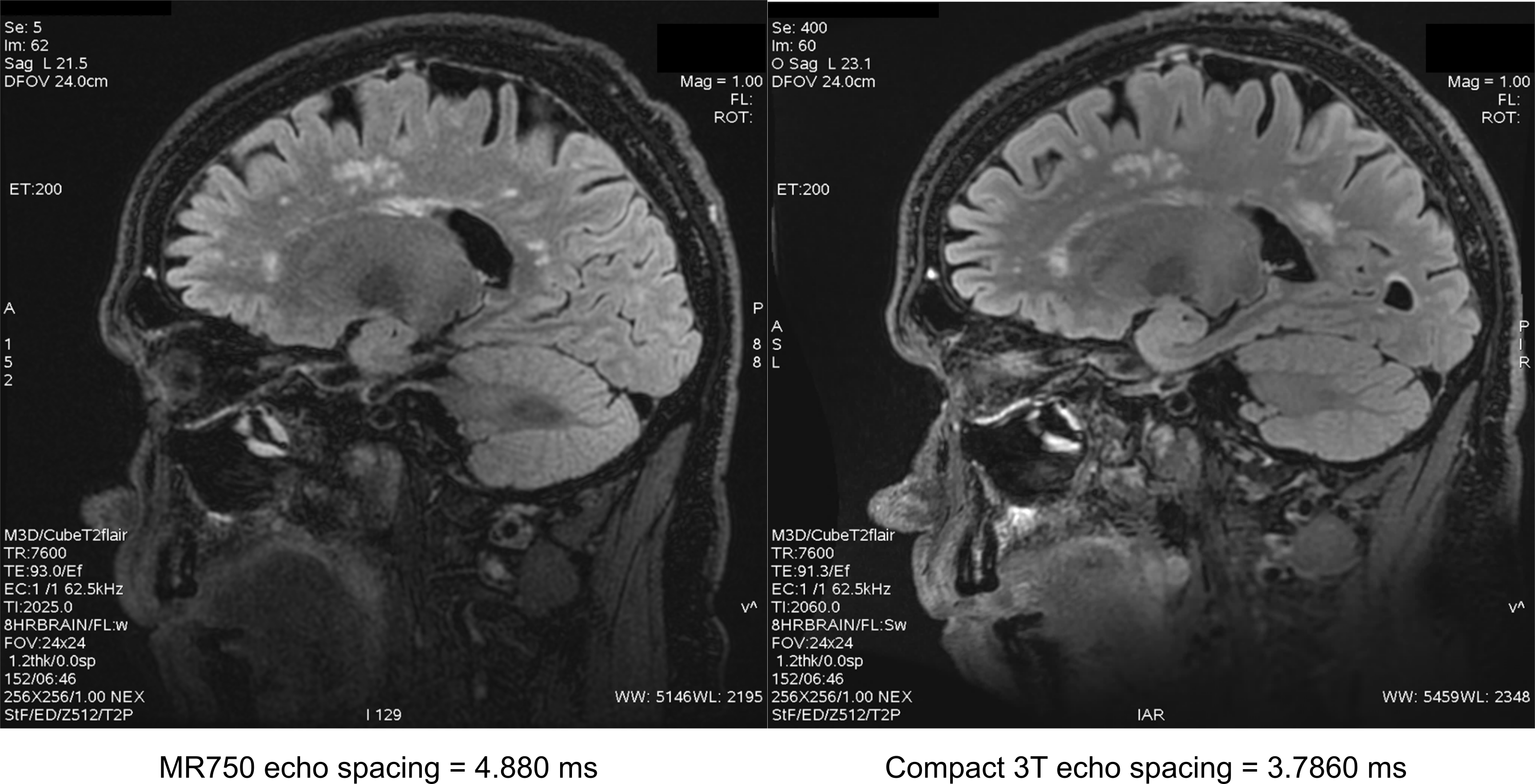
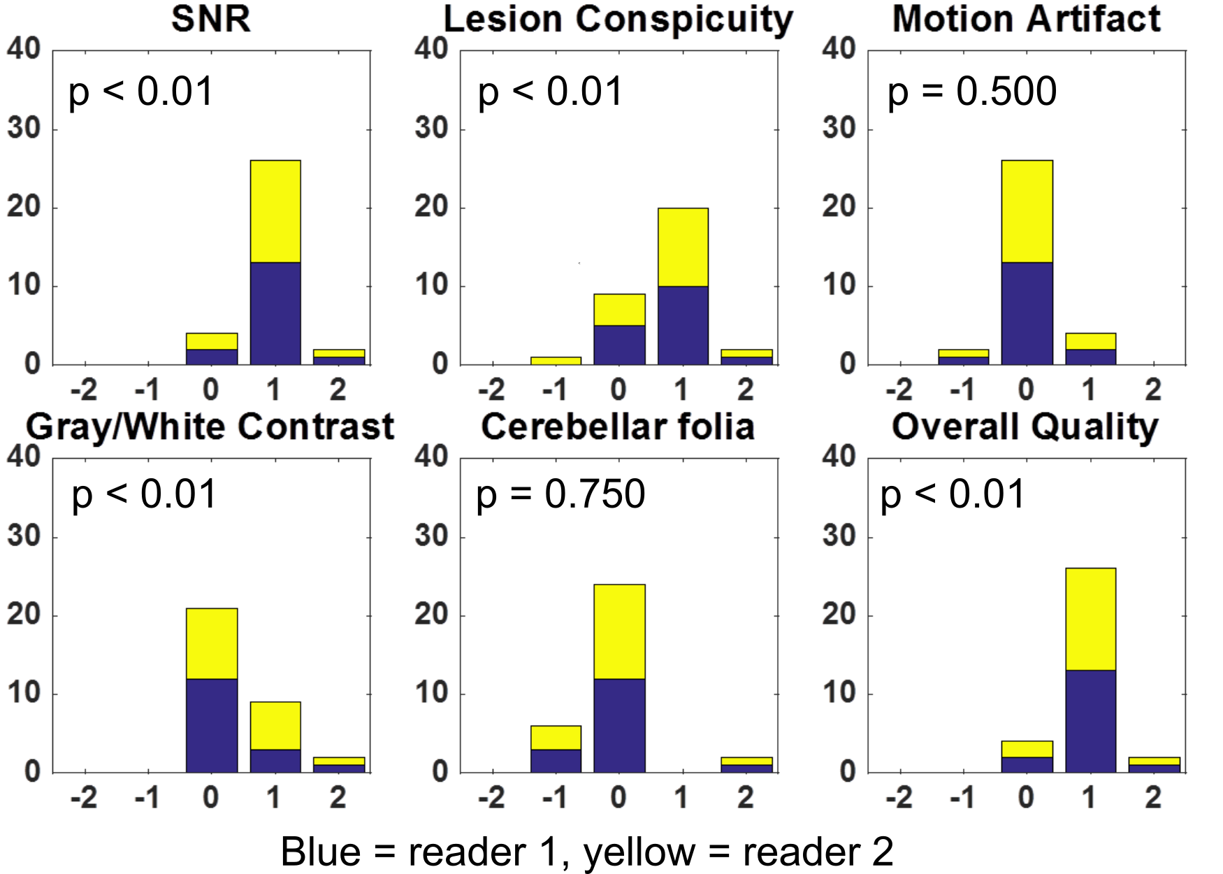
Methods: Under an IRB-approved protocol, 16 clinical patients receiving a head MR examination were scanned both on a standard whole-body MR system (GE Discovery MR750, GE Healthcare, Waukesha, WI) and the compact 3T using an 8-channel receiver coil (In-vivo, Cleveland, Ohio). The sagittal T2-weighted 3D FLAIR (i.e. Cube-Flair) parameters were as follows: 256 × 256 × 152 (frequency × phase × partition) matrix, 24 × 24 × 18.2 cm field-of-view (FOV), 7600 ms repetition time (TR), 93.0 ms effective echo time (TE) and 2025 ms inversion time (TI) on the whole body system, 91.3 ms TE and 2060 ms TI on the compact system, an echo-train-length of 200, with an echo spacing (ESP) of 4.880 ms on the whole body and 3.768 ms on the compact system. The compact 3T used real-time gradient pre-emphasis[4] and frequency shifting to compensate additional concomitant fields. Both exams used a self-calibrating data-driven parallel imaging (ARC)[5] acceleration factor of R = 2. The exams were reviewed by two board-certified neuroradiologists with 17 and 26 years of experience. They were graded on a five-point ordinal scale from -2 to +2, with +2 indicating strong preference for the compact system and -2 indicating strong preference for the whole-body system, +1 and -1 indicating slight preference for the compact and whole body systems respectively, and 0 representing no system preference. Each pair of images was comparatively evaluated (non-blinded) using the following attributes: signal-to-noise ratio (SNR), lesion conspicuity, motion artifact, gray/white matter contrast, cerebellar folia conspicuity, and overall exam quality. The sample size of 16 was chosen to enable detection of a 10% scoring difference in like uncertainty using a one-sided Wilcoxon signed rank tests with 80% power (efficiency adjusted[6]) at a 5% significance level. The null hypothesis of the left-sided test was that the compact 3T performed equally to or better than the standard whole-body 3T MR system; and the right-sided test used the reverse of this hypothesis. Krippendorf’s Alpha[7] (ordinal difference) was computed for the pooled score sets to characterize inter-reader agreement.Results
Figure 1 shows a representative comparison between images from the two scanners. This example shows highlights a patient with small vessel ischemic disease. The sagittal image demonstrates the typical white matter T2 hyperintensities associated with the ischemic degenerative changes of aging.. The results of the radiologic evaluation are shown in Figure 2, with pooled histograms of each of the six criteria shown. As strong inter-reader agreement was observed (α=0.92), all testing was performed on the inter-reader average scores. The significance results of the right-tailed test are overlaid onto these histograms. Presuming a 5% significance level, the right-sided test results indicate that the compact 3T outperformed the whole body system in terms of SNR, lesion conspicuity, gray/white matter contrast, and overall quality. All left-sided tests also failed to reject the null hypothesis, indicating that the compact 3T system performed as good as the whole-body system with respect to motion artifact and cerebellar folia conspicuity.Discussion
There is strong statistical evidence that the compact 3T provides equal or better image quality with the 3D FLAIR sequence across this sample set. The unchanged incidence of motion artifact also suggests that the compact system offers at least equivalent patient comfort, while high slew-rate gradient system has allowed for an ESP reduction of 25% compared to the whole-body system. Over a long ETL sequence like 3D FSE, this reduction yields an overall readout duration change from 960 ms to 747 ms, allowing for the acquisition of higher spatial frequencies with less T2 signal decay.Conclusion
The compact 3T provided equal or better image quality compared to a standard whole-body 3T for a standard clinical imaging sequence across a small patient cohort.Acknowledgements
This work was supported in part by NIH grant RO1EB010065.References
1] Lee S-K, Mathieu J-B, Graziani D, Piel J, Budesheim E, Fiveland E, et al. Peripheral nerve stimulation characteristics of an asymmetric head-only gradient coil compatible with a high-channel-count receiver array. Magn Reson Med 2015:n/a-n/a. doi:10.1002/mrm.26044.
[2] Mathieu J-B, Lee S-K, Graziani D, Lin J, Budesheim E, Piel JE, et al. Development of a Dedicated Asymmetric Head-only Gradient Coil for High-Performance Brain Imaging with a High PNS Threshold. ISMRM Annu. Meet., Toronto: 2015, p. 1019.
[3] Weavers PT, Shu Y, Tao S, Huston J, Lee S-K, Graziani D, et al. Technical Note: Compact three-tesla magnetic resonance imager with high-performance gradients passes ACR image quality and acoustic noise tests. Med Phys 2016;43:1259–64. doi:10.1118/1.4941362.
[4] Tao S, Weavers PT, Trzasko JD, Shu Y, Huston J, Lee S, et al. Gradient pre-emphasis to counteract first-order concomitant fields on asymmetric MRI gradient systems. Magn Reson Med 2016;0. doi:10.1002/mrm.26315.
[5] Brau ACS, Beatty PJ, Skare S, Bammer R. Comparison of reconstruction accuracy and efficiency among autocalibrating data-driven parallel imaging methods. Magn Reson Med 2008;59:382–95. doi:10.1002/mrm.21481.
[6] Siegel S. Nonparametric Statistics for the Behavioral Sciences. McGraw-Hill; 1956.
[7] Hayes AF, Krippendorff K. Answering the Call for a Standard Reliability Measure for Coding Data. Commun Methods Meas 2007;1:77–89. doi:10.1080/19312450709336664.
Figures