4739
Parallel transmission (pTx) on an in-vivo human brain at 7T: a new approach using global B1 coefficient template1Imaging Institute, Cleveland Clinic Foundation, Cleveland, OH, United States, 2Department of Biomedical Engineering, Hankuk University of Foreign Studies, Yongin, Korea, Republic of, 3Siemens Healthcare, New York, NY, United States
Synopsis
Improving B1-uniformity by calibration procedure is crucial for the pTx imaging and commonly individual B1 coefficient estimation is conducted in the beginning of each patient scan protocol. However, B1 calibration procedure increase the scan time by approximately 10~15 mins increasing discomfort to the patient. Here we propose using global B1 coefficient template to reduce scan time while maintaining acceptable B1 profile. The approach using global B1 coefficient template is assumed to have limited inter-individual variation of B1 coefficient between subjects. We demonstrate the feasibility of scan with global B1 coefficient template and provide a perception of in-vivo scan with pTx.
Introduction
MR imaging at ultra-high fields (UHF) is becoming increasingly common in the neuroimaging research field. As the magnetic field strength increases, net magnetization also increases, leading to a higher signal-to-noise ratio (SNR) and allowing for high-resolution MR imaging. UHF leads to sub-millimeter anatomic imaging in a clinically reasonable scan time and has helped to correct misinterpretations of scans performed on lower fields1. High-resolution UHF MR imaging is much helpful when the surgical lesions are so small. Moreover, surgical areas can be minimized with high-resolution MR images. However, problems at UHF, most notably B1-field inhomogeneity induced signal distortion, limit patient studies. These problems are magnified in the lower temporal lobe.
Parallel transmission MRI (pTx), utilized with multiple-transmit coil elements, was proposed to address the aforementioned non-uniform B1 issue particularly at UHF2. Theoretically, if the B1 shimming parameters were accurately estimated pTx can provide uniform slice-select excitation and improve spatially dependent tissue contrast. Therefore, improving B1-uniformity by calibration procedure is crucial part for the pTx imaging and commonly individual B1 coefficient estimation is conducted in the beginning of each patient scan protocol. However, B1 calibration procedure increase the scan time by approximately 10~15 mins increasing discomfort to the patient.
Here we propose using global B1 coefficient template to reduce scan time while maintaining acceptable B1 profile. The approach using global B1 coefficient template is assumed to have limited inter-individual variation of B1 coefficient between subjects. We demonstrate the feasibility of scan with global B1 coefficient template and provide a perception of in-vivo scan with 7T pTx system.Methods
To investigate the inter-individual variation of the B1 coefficient, two healthy controls data were acquired with 7T pTx (step 2.3) MRI scanner (IRB-approved, Siemens). An 8-channel transmit and 32-channel receive phased array coil (Nova Medical) was used. 3 different B1 (amplitude and phase) calibration parameters (CP, N1 and N2) were estimated from the first subject (Sub #1) using scanner embedded tool. CP mode and N1 were optimized for temporal pole and cerebellum imaging, respectively. N2 has intermediate value between CP and N1. And these B1 parameters were applied for both (Sub #1 and #2) volunteer scans. The scan parameters for MPRAGE were as follows: TR/TE/TI = 4000/1.8/2300 ms, flip angle = 8°, 192 slices, 0.9 mm3-isotropic voxel, GRAPPA factor = 3 and 4.4 min.Results
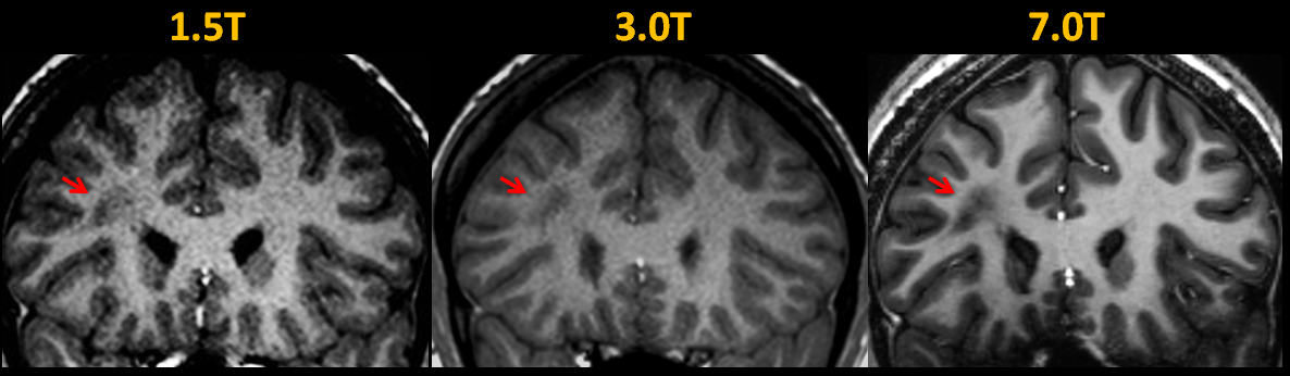
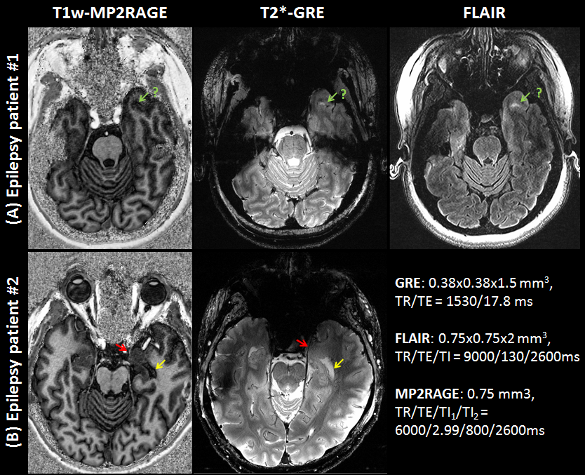
Figure 1 shows epilepsy patient images obtained using 3 different modalities. Image acquired with higher-field-strength clearly showed the lesion. As examples of clinical applications, Fig. 2 features a 7T images preoperatively obtained from patients with epilepsy. Due to signal loss and non-uniform contrast at lower temporal lobe, the lesions are not clearly shown.
Figure 3 displays the results of the global B1 coefficient template. When compared, both subject data show similar spatial distributions of the signal and contrast. In terms of gray and white matter tissue contrast, results with CP mode show improved contrast at the temporal pole region but degenerated contrast at the cerebellum. Compared to the scan with CP mode B1 parameter, results with N1- and N2-B1 parameters show opposite and intermediate values.
Discussion and Conclusion
The image quality in sTx system has been much improved by using less B1 sensitive sequence such as MP2RAGE3. However, low sensitivity and non-uniform contrast in the temporal pole area still has been a challenge. In this work, a new approach using global B1 coefficient template for the 7T pTx scan is demonstrated. The experimental results show that the variation of the calibrated B1 parameters between subjects is limited, confirming that the use of global B1 coefficient template can be acceptable. Proposed approach can be shorten the scan time as well as reduced discomfort to the patient. Based on these observations, B1 coefficient templates estimated only for the specific brain area (i.e. temporal pole, cerebellum) might be useful for the patient scan with pTx system. This is an initial study of 7T pTx in-vivo anatomic scanning. This study serves as a starting point toward pTx in-vivo clinical scanning. In addition, it gives some insight as to current challenges and future work.Acknowledgements
This work was supported by Cleveland Clinic. Author gratefully acknowledges technical support by Siemens Medical Solutions. Authors thank Dr. Tobias Kober for supporting MP2RAGE WIP.References
[1] Stephen E Jones, Se-Hong Oh, Erik Beall, Michael Phillips, Ken Sakaie, Irene Wang, and Mark Lowe, “Examples of clinical imaging at 7T: Successes and Challenges”, ISMRM, 2015, 767
[2] Yudong Zhu, “Parallel excitation with an array of transmit coils”, MRM, 2004, 51(4):775-84
[3] Marques JP, Kober T, Krueger G, van der Zwaag W, Van de Moortele PF, Gruetter R. “MP2RAGE, a self bias-field corrected sequence for improved segmentation and T1-mapping at high field.”, Neuroimage, 2010, 49(2):1271-81
Figures