4736
A method for near-realtime automated segmentation of thalamic nuclei1Electrical and Computer Engineering, Univ. of Arizona, Tucson, AZ, United States, 2Radiology, Stanford University, Stanford, CA, United States, 3Medical Imaging, Univ. of Arizona, Tucson, AZ, United States
Synopsis
Thalamic nuclei are often hard to visualize on most anatomical sequences. White-matter-nulled MPRAGE imaging provides sufficient intra-nuclear contrast to enable manual segmentation, which is very tedious. We have developed fast multi-atlas based segmentation schemes that can provide accurate segmentation of all the major thalamic nuclei in under 15 minutes.
Introduction
Precise localization and volumetry of thalamic nuclei are critical for deep brain surgery (DBS) targeting and understanding the effect of Alzheimer’s, schizophrenia and other pathologies on specific thalamic nuclei. To date, MRI based methods have been suboptimal. Recently, a white-matter-nulled MP-RAGE sequence along with manual [1] or automated [2-3] segmentation has shown considerable promise, despite long processing times. We report on a novel, fast technique that has the potential for near-realtime segmentation of thalamic nuclei, achieving up to 16x speedup over current methods.Methods
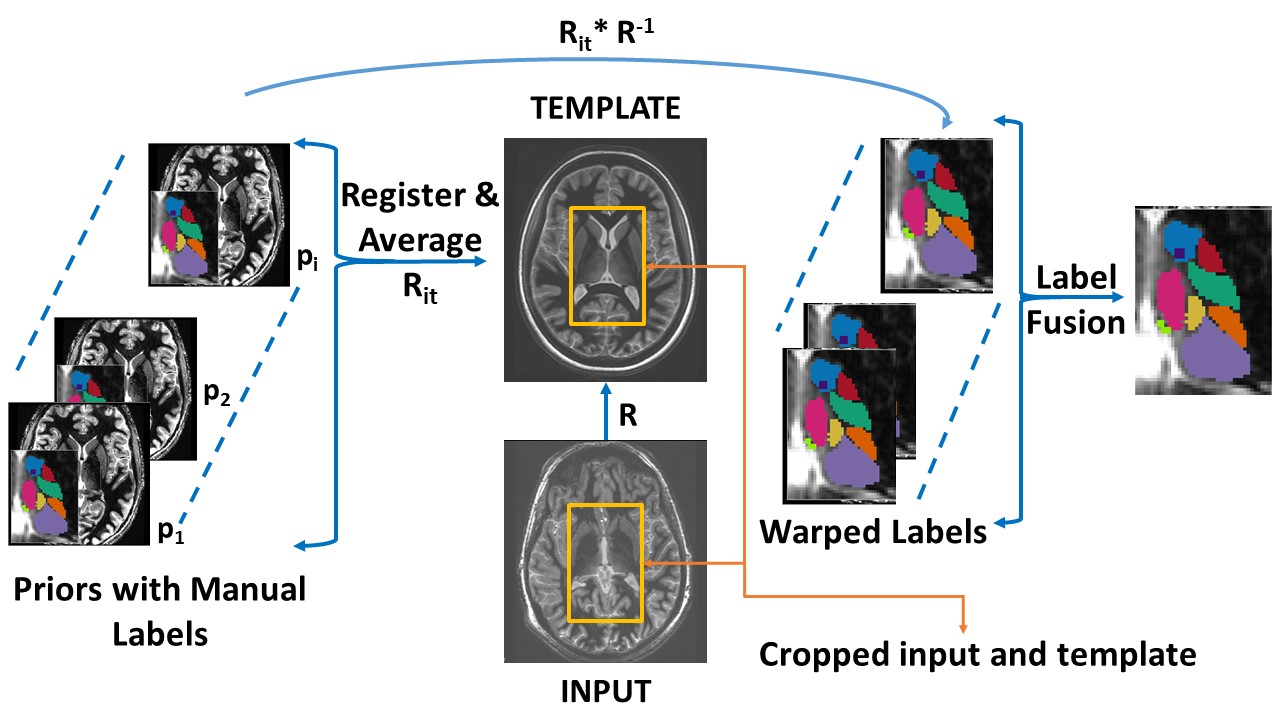
Technique: Two variants of THalamus Optimized Multi-Atlas Segmentation (THOMAS) have been proposed [2-3]. Both use a “multi-atlas” comprising of 20 white-matter-nulled MPRAGE prior datasets (12 patients and 8 controls) with manual segmentation by a neuroradiologist [1], guided by the Morel atlas. We developed a fast and robust hybrid of these two methods (Fig 1), using an intermediary averaged template for robustness and cropping the template and input from for speed. The crop size was varied systematically from full size to the smallest crop that encompasses both thalami in all 3 dimensions. Further speedup was achieved using Majority voting (MV) instead of Joint fusion (JF), for label fusion. We hypothesized that the robustness of template based registration would enable the use of MV label fusion, significantly reducing processing times whilst maintaining accuracy. We call our new method ST THOMAS (Shortened Template and THalamus for Optimal Multi Atlas Segmentation).
The two THOMAS variants and ST THOMAS were compared on 20 subjects (12 patients with MS) who were scanned after informed consent on a 7T GE MRI scanner using a white-matter-nulled MP-RAGE pulse sequence. THOMAS-1 was implemented using joint fusion, a more robust algorithm than STEPS label fusion of [2]. Even though 11 nuclei were generated, the 5 nuclei used for analysis in addition to the whole thalamus (Thal) were ventral lateral (VL), mediodorsal (MD), pulvinar (Pul), anterior ventral (AV) and ventral lateral posterior (VLP). We used processing times, Dice Similarity Coefficients (DSC) and Volume Similarity Index (VSI) as quantitative metrics for comparison. DSC is a measure of overlap accuracy whilst VSI measures accuracy of estimated volumes. All experiments were conducted on a dual-CPU 4-core 3 GHz Intel Xeon E5-2623 v3 Dell workstation.
Results
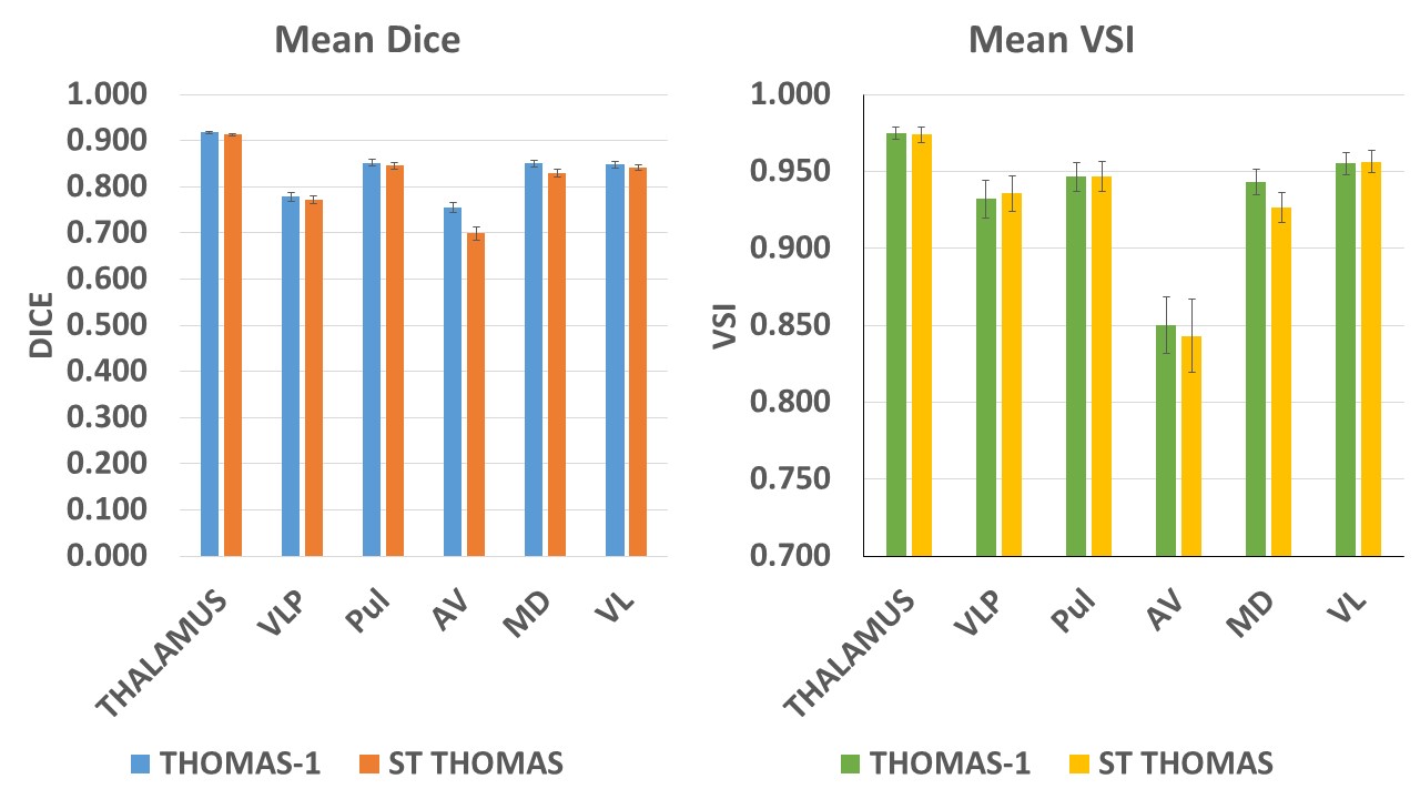
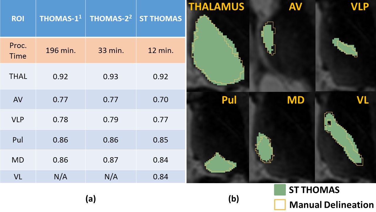
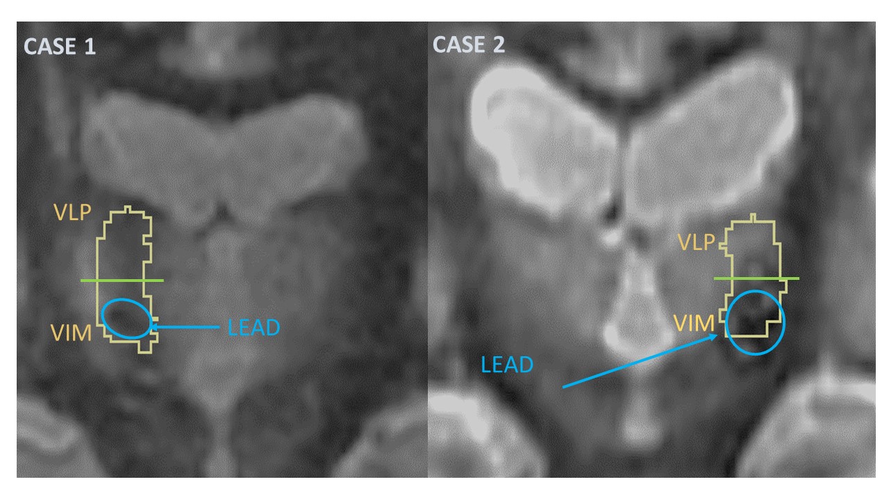
The proposed method ST THOMAS had mean processing times of 66 min and 10 min respectively for Joint fusion and Majority voting. The corresponding times for THOMAS-1 were 196 and 134 min respectively. Mean processing times for THOMAS-2 with joint fusion was 30 min. Figure 2 shows mean processing times and Dice indices for different thalamic nuclei as a function of crop size for our proposed method using MV. It can be seen that the shortest processing times (7 and 12 min.) are achieved for the two smallest crop sizes. Since the Dice indices for 3 nuclei (AV, VLP, Pul) were slightly higher (p < 0.05) for the second smallest crop (93x187x68), we used that for the rest of the analysis. Figure 3 compares ST THOMAS with mean processing time of 12 min and THOMAS-1 with 196 min mean processing time for Dice and VSI. The results are very comparable with the worst performing nucleus for ST THOMAS, 7 % lower Dice for AV and 1.7 % lower VSI for MD. Note that the VLP nucleus whose inferior part or the ventral intermediate (VIM) nucleus and is critical for DBS targeting is comparable in accuracy for ST THOMAS (p=0.3). Table 1 compares the performance (median Dice) of ST THOMAS with the two methods reported in literature [2-3]. It can be seen that we have achieved comparable accuracy whilst achieving 16X and 3X reduction in processing times over the two methods. Figure 4 shows segmented VLP nucleus from two pre-operative DBS patients overlaid after registration on post-operative MP-RAGE images. In both cases, the inferior part of the VLP nucleus (which is the VIM) corresponds well with the DBS lead (black dephased area), suggesting the feasibility of ST THOMAS for realtime DBS surgery.Conclusions
Our novel segmentation pipeline provides a new baseline in the work towards automated segmentation of thalamic nuclei with increased speedup (3X-16X) and high accuracy. By combining optimal cropping of the template and input as well as using majority voting (as opposed to joint fusion), we can achieve 12 min. processing times, opening the avenue for realtime DBS surgery applications.Acknowledgements
References
1. Tourdias et al. Neuroimage. 2013 Sep 7;84C:534-545
2. Su et al. Proceedings of ISMRM. 2014 May 10;4306
3. Su et al. Proceedings of ISMRM. 2015 May 30;6231
Figures