4726
A Retrospective Study of Amide Proton Transfer Imaging in Acute Ischemic Stroke1Department of Radiology, Beijing Hospital, Beijing, People's Republic of China, 2Department of Radiology, Johns Hopkins University, BALTIMORE, MD
Synopsis
This study was performed to investigate the imaging features of Amide Proton Transfer (APT) MR imaging in different subtypes of acute ischemic stroke (AIS) . By figuring out the detailed APTw features in AIS patients, it would be helpful for further clinical applications of APT MRI technique.
Purpose: This study was performed to investigate the imaging features of Amide Proton Transfer (APT) MR imaging in different types of acute ischemic stroke (AIS).
Methods: 93 clinically diagnosed AIS subjects presenting within 72 hours of symptom onset were enrolled. APT and routine MR imaging were performed in all subjects. Patients were divided into four subtypes defined by the radiological Oxfordshire Community Stroke Project (OCSP) classification: total anterior circulation infarcts (TACI, n = 9), partial anterior circulation infarcts (PACI, n = 28), lacunar circulation infarcts (LACI, n = 39), and posterior circulation infarcts (POCI, n = 17). APT weighted (APTw) signal intensities in the infarcted tissue (APTWlesion) and in the contralateral normal-appearing region (APTWcontral) were measured. Two-sample t test and one-way ANOVA was performed to evaluate the differences of APTw signal intensities in the four subtypes.
Results: Of all enrolled patients, the overall average APTw signal intensities were lower in the ischemic tissue than the control normal brain tissue, corresponding to (- 0.01 ± 0.81) % and (0.62 ± 0.44) %, respectively (P < 0.001). APTWlesion values were (0.51 ± 0.66) % in the LACI group, (- 0.38 ± 0.59) % in the PACI group, (- 0.02 ± 0.76) % in the POCI group, and (- 1.01 ± 0.33) % in the TACI group. Subtype analysis showed that there was no significantly difference between APTWlesion and APTWcontral values (P = 0.072) in the LACI group. However, the differences between APTWlesion and APTWcontral were significant in the TACI, PACI and POCI group (P < 0.05 in all the three groups). Patients with TACI had the lowest APTWlesion values than other subtypes. APTWcontral values showed no significant differences among the four groups. Patients with POCI showed no significant differences between APTWlesion and APTWcontral in patients with brainstem and cerebellum stroke (P=0.550).
Conclusion: APT MRI can sensitively detect the existence of acidosis in AIS and distinguish the difference between the infarcted tissue and the normal regions in TACI and PACI patients. However, Patients with LACI and POCI would not benefit much from APT MRI.
Acknowledgements
This work was supported in part by grants from the National Natural Science Foundation of China (81361120392 and 81401404), Beijing Natural Science Foundation (7154235), and the National Institutes of Health (R01NS083435, R01EB009731, and R01CA166171).References
1. Zhou JY, Payen JF, Wilson DA, Traystman RJ, van Zijl PCM. Using the amide proton signals of intracellular proteins and peptides to detect pH effects in MRI. Nat Med. 2003;9(8):1085-90.
2. Zhou J, Lal B, Wilson DA, Laterra J, van Zijl PC. Amide proton transfer (APT) contrast for imaging of brain tumors. Magn Reson Med. 2003;50(6):1120-6.
3. Harston GWJ, Tee YK, Blockley N, Okell TW, Thandeswaran S, Shaya G et al. Identifying the ischaemic penumbra using pH-weighted magnetic resonance imaging. Brain. 2015;138:36-42.
4. Tietze A, Blicher J, Mikkelsen IK, Ostergaard L, Strother MK, Smith SA et al. Assessment of ischemic penumbra in patients with hyperacute stroke using amide proton transfer (APT) chemical exchange saturation transfer (CEST) MRI. NMR Biomed. 2014;27(2):163-74.
5. Zhou JY, van Zijl PCM. Defining an Acidosis-Based Ischemic Penumbra from pH-Weighted MRI. Transl Stroke Res. 2012;3(1):76-83.
6. Sun PZ, Cheung JS, Wang E, Lo EH. Association between pH-weighted endogenous amide proton chemical exchange saturation transfer MRI and tissue lactic acidosis during acute ischemic stroke. J Cereb Blood Flow Metab. 2011;31(8):1743-50.
Figures
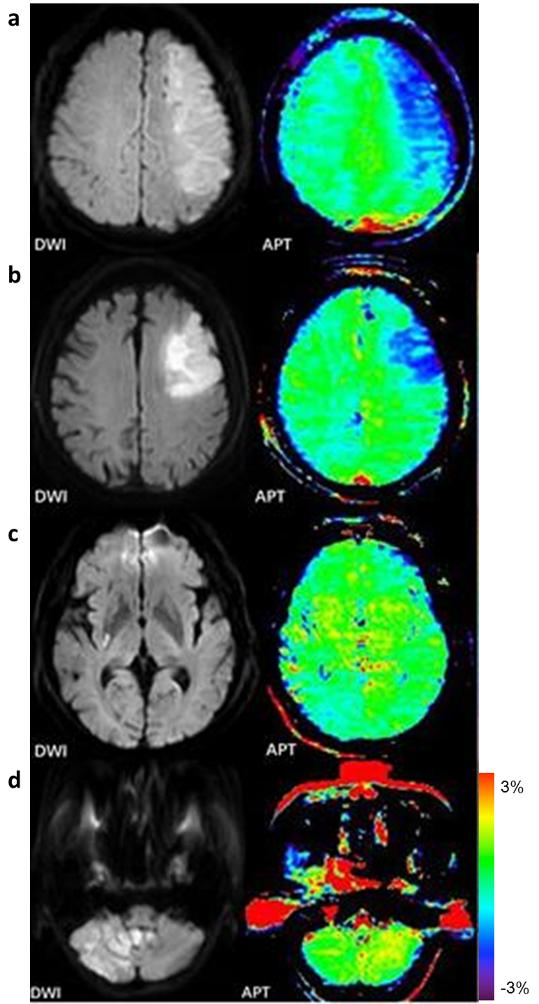
Four patients in different subtypes.
a: a patient with TACI. F/50y, APTWlesion=-1.44%, APTWcontral=0.22%.
b: a patient with PACI. APTWlesion= -0.80%, APTWcontral= 0.47%.
c: a patient with LACI. APTWlesion=1.07%, APTWcontral=1.25%.
d: a patient with POCI. APTWlesion= 0.48%, APTWcontral=1.66%.