4720
Scheme optimization for inflow and outflow visualization in non-contrast enhanced dynamic MRA based on pseudo-continuous arterial spin labeling1Philips Electronics Japan, Tokyo, Japan, 2Clinical Radiology, Graduate School of Medical Sciences, Kyushu University, Fukuoka, Japan, 3Department of Radiology, Hokkaido University Hospital, Hokkaido, Japan, 4Department of Radiology, Tokai University Hospital, Kanagawa, Japan, 5Asia Pacific, Philips Healthcare, Tokyo, Japan
Synopsis
A new scheme for non-contrast enhanced intracranial three-dimensional dynamic magnetic resonance angiography (4D-MRA) using pseudo-continuous arterial spin labeling (4D-PCASL) is proposed for visualizing inflow and outflow dynamics. The 4D-PCASL procedure was accelerated with contrast-enhanced timing-robust angiography (CENTRA)-Keyhole and the view-sharing techniques (4D-PACK). Images acquired from four volunteers were compared between the 4D-PCASL and 4D-PACK approaches. We show that this new scheme accelerates data acquisition and provides dynamic inflow and outflow information.
INTRODUCTION
Arterial spin labeling (ASL) has a demonstrated capacity for non-contrast enhanced intracranial three-dimensional (3D) dynamic magnetic resonance angiography (4D-MRA).1-15 One of the approaches for 4D-MRA is to use pseudo-continuous ASL (4D-PCASL), which provides a high flow signal.3,6,8,10 One drawback to the 4D-PCASL procedure is scan time prolongation. A solution proposed recently, termed 4D-PACK, accelerates acquisition using contrast-enhanced timing-robust angiography (CENTRA)-Keyhole and view-sharing techniques.8 There are two schemes to observe dynamic information in 4D-PCASL. One is to increase labeling duration (LD)3,8, which is termed the 4D-PCASL-LD and the other is to increase post labeling delay (PLD)6,10, which is termed the 4D-PCASL-PLD later on. The 4D-PCASL-LD technique is expected to be advantageous in peripheral artery visualization, because it utilizes a long LD. On the other hand, it visualizes only inflow dynamics and cannot observe outflow dynamics. In 4D-PCASL-PLD, both inflow and outflow dynamics can be observed if the LD is sufficiently short. However, a short LD can lead to insufficient vessel visualization. To observe both inflow and outflow dynamics with sufficient visualization, we propose the 4D-PCASL-Hybrid scheme, combining 4D-PCASL-LD and 4D-PCASL-PLD. In this study we implemented the 4D-PCASL-Hybrid sequence and further combined it with the 4D-PACK acceleration (4D-PACK-Hybrid). The aim of this study is to assess the inflow and outflow dynamic information of the Hybrid approach and to compare 4D-PCASL-Hybrid with 4D-PACK-Hybrid.METHODS
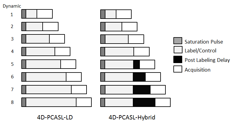
4D-PCASL-Hybrid scheme The 4D-PCASL-LD and 4D-PCASL-Hybrid schemes are shown in Figure 1. Inflow dynamic data can be acquired by changing the LD in the 4D-PCASL-LD. In the 4D-PCASL-Hybrid, inflow dynamics are acquired in the first half of the dynamics by increasing the LD, similar to the 4D-PCASL-LD scheme, and acquiring the outflow dynamics by changing the PLD in second half of the dynamics.
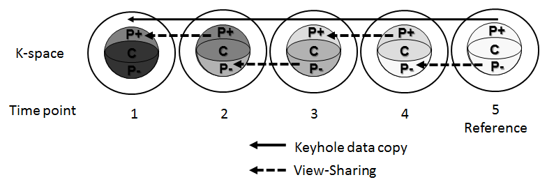
4D-PACK The overall 4D-PACK steps are described in Figure 2. Only the elliptical central disk data are sequentially acquired along with the time points except at the reference point.16 In the Keyhole technique, the missing profiles in the k-space of the high frequency region were complemented by the reference data. In addition, the view-sharing technique was combined. In this technique, the central disk is subdivided into three regions, P+, C and P-, as shown in Figure 2.17 While the central region C is acquired at every time point, the peripheral regions P+ and P- are acquired in an alternating fashion, and missing profiles are matched with subsequent time points.
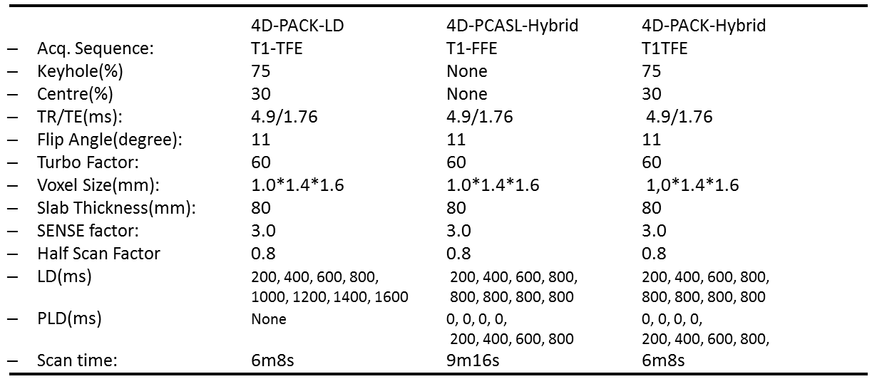
Magnetic Resonance (MR) Experiments The 4D-PACK-Hybrid scheme was implemented on a 3.0T scanner (Philips Ingenia R5). Four healthy subjects were examined after obtaining informed consent as required by the Institutional Review Board. The 4D-MRA data were acquired for comparison by three sequences, the 4D-PACK-LD, 4D-PCASL-Hybrid, and 4D-PACK-Hybrid. Acquisition parameters for each sequence are summarized in Table 1. For image evaluation of the 4D-PACK-LD, 4D-PCASL-Hybrid, and 4D-PACK-Hybrid, axial maximum intensity projection (MIP) images were generated with a full volume thickness of 80 mm at each time point. A circular region of interest (ROI) was selected from the white matter (WM) and the middle cerebral artery (MCA). Multiple ROIs were placed on the left and right MCA regions through the M1, M2, M3, and M4 branches.
Validation of flow dynamic information For validation of flow dynamic information, inflow time (IT) and outflow time (OT) were measured in the 4D-PCASL-Hybrid and 4D-PACK-Hybrid at multiple regions in the MCA. For IT and OT measurements, the maximum signal within each ROI was measured at each time point. Then, extrapolated linearly from the signals at each time point, a time intensity curve at each ROI in the MCA was calculated. Here, the IT and OT were defined as the time points for the signal to reach its half maximum value before and after the signal peak, respectively.5,18 We then measured the correlation coefficient and slope of approximation for the line between the ITs and OTs in the 4D-PCASL-Hybrid and 4D-PACK-Hybrid.
RESULTS
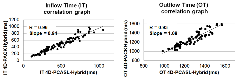
The representative dynamic data acquired by the 4D-PACK-LD, 4D-PCASL-Hybrid, and 4D-PACK-Hybrid are shown in Figure 3. The Hybrid schemes provide both inflow and outflow dynamic information, while the LD scheme provides only inflow dynamics. The IT and OT correlation coefficients between the 4D-PCASL-Hybrid and 4D-PACK-Hybrid were determined to be 0.96 (IT) and 0.93 (OT) as shown in Figure 4. The slopes of the predicted lines were 0.94 (IT) and 1.08 (OT).CONCLUSIONS
The 4D-PCASL-Hybrid scheme provides both inflow and outflow dynamic information. In addition, it can be accelerated by using the CENTRA-Keyhole and view-sharing technique. This accelerated 4D-PACK-Hybrid sequence retains the dynamic information.Acknowledgements
We thank Tetsuo Ogino for valuable help with the imaging sequence development and Yuta Akamine for checking the abstract contents.References
1. Yu S, Yan L, Yao Y, et al. Noncontrast dynamic mra in intracranial arteriovenous malformation (avm), comparison with time of flight (tof) and digital subtraction angiography (dsa). Magn Reson Imaging 2012;30:869-877.
2. Yan L, Wang S, Zhuo Y, et al. Unenhanced dynamic mr angiography: high spatial and temporal resolution by using true fisp-based spin tagging with alternating radiofrequency. Radiology 2010;256:270-279.
3. Wu H, Block WF, Turski PA, et al. Noncontrast dynamic 3d intracranial mr angiography using pseudo-continuous arterial spin labeling (pcasl) and accelerated 3d radial acquisition. J Magn Reson Imaging 2014;39:1320-1326.
4. Tan ET, Huston J 3rd, Campeau NG, et al. Fast inversion recovery magnetic resonance angiography of the intracranial arteries. Magn Reson Med 2010;63:1648-1658.
5. Robson PM, Dai W, Shankaranarayanan A, et al. Time-resolved vessel-selective digital subtraction mr angiography of the cerebral vasculature with arterial spin labeling. Radiology 2010;257:507-515.
6. Okell TW, Schmitt P, Bi X, et al. Optimization of 4d vessel-selective arterial spin labeling angiography using balanced steady-state free precession and vessel-encoding. NMR Biomed 2016;29:776-786.
7. Okell TW, Chappell MA, Woolrich MW, et al. Vessel-encoded dynamic magnetic resonance angiography using arterial spin labeling. Magn Reson Med 2010;64:430-438.
8. Obara M, Togao O, Okuaki T, et al. Non-contrast enhanced 4d intracranial mr angiography based on pseudo-continuous arterial spin labelling (pcasl) with the keyhole technique. In:Proc 24th Annual Meeting of ISMRM, Singapore 2016;:4376.
9. Nakamura M, Yoneyama M, Okuaki T, et al. Non contrast 3d volumetric time-resolved mra combining multiple phase fair(cinema-fair). In:Proc 19th Annual Meeting of ISMRM, Montreal, Canada 2011;:4036.
10. Lindner T, Jensen-Kondering U, van Osch MJ, et al. 3d time-resolved vessel-selective angiography based on pseudo-continuous arterial spin labeling. Magn Reson Imaging 2015;33:840-846.
11. Iryo Y, Hirai T, Nakamura M, et al. Collateral circulation via the circle of willis in patients with carotid artery steno-occlusive disease: evaluation on 3-t 4d mra using arterial spin labelling. Clin Radiol 2015;70:960-965. 12. Iryo Y, Hirai T, Kai Y, et al. Intracranial dural arteriovenous fistulas: evaluation with 3-t four-dimensional mr angiography using arterial spin labeling. Radiology 2014;271:193-199.
13. Fujima N, Osanai T, Shimizu Y, et al. Utility of noncontrast-enhanced time-resolved four-dimensional mr angiography with a vessel-selective technique for intracranial arteriovenous malformations. J Magn Reson Imaging 2016;44:834-845.
14. Bi X, Weale P, Schmitt P, et al. Non-contrast-enhanced four-dimensional (4d) intracranial mr angiography: a feasibility study. Magn Reson Med 2010;63:835-841. 15. Uchino H, Ito M, Fujima N, et al. A novel application of four-dimensional magnetic resonance angiography using an arterial spin labeling technique for noninvasive diagnosis of moyamoya disease. Clin Neurol Neurosurg 2015;137:105-111.
16. Willinek WA, Gieseke J, Conrad R, et al. Randomly segmented central k-space ordering in high-spatial resolution contrast-enhanced mr angiography of the supraaortic arteries: initial experience. Radiology 2002;225:583-588.
17. Hadizadeh DR, Gieseke J, Beck G, et al. View-sharing in keyhole imaging: partially compressed central k-space acquisition in time-resolved mra at 3.0 t. Eur J Radiol 2011;80:400-406.
18. Riederer SJ, Haider CR, Borisch EA. Time-of-arrival mapping at three-dimensional time-resolved contrast-enhanced mr angiography. Radiology 2009;253:532-542.
Figures